中国寄生虫学与寄生虫病杂志 ›› 2022, Vol. 40 ›› Issue (1): 28-35.doi: 10.12140/j.issn.1000-7423.2022.01.004

• 论著 • 上一篇    下一篇

高剂量氯磷酸脂质体处理对小鼠体内约氏疟原虫生长的影响及机制初探

陈穗林(), 高源利, 郭帅, 范永铃, 刘太平, 徐文岳*()   

  1. 陆军军医大学基础医学院病原生物学教研室,重庆 400038
  • 收稿日期:2021-12-02 修回日期:2021-12-21 出版日期:2022-02-28 发布日期:2022-01-24
  • 通讯作者: 徐文岳
  • 作者简介:陈穗林(1997-),男,硕士研究生,从事疟疾感染免疫研究。E-mail: chensuilin0208@163.com
  • 基金资助:
    国家自然科学基金面上项目(82172296)

Effect and mechanism of high-dose clodronate liposomes treatment on Plasmodium yoelii growth in mice

CHEN Sui-lin(), GAO Yuan-li, GUO Shuai, FAN Yong-ling, LIU Tai-ping, XU Wen-yue*()   

  1. Department of Pathogenic Biology, Army Medical University, Chongqing 400038, China
  • Received:2021-12-02 Revised:2021-12-21 Online:2022-02-28 Published:2022-01-24
  • Contact: XU Wen-yue
  • Supported by:
    National Natural Science Foundation of China(82172296)

摘要:

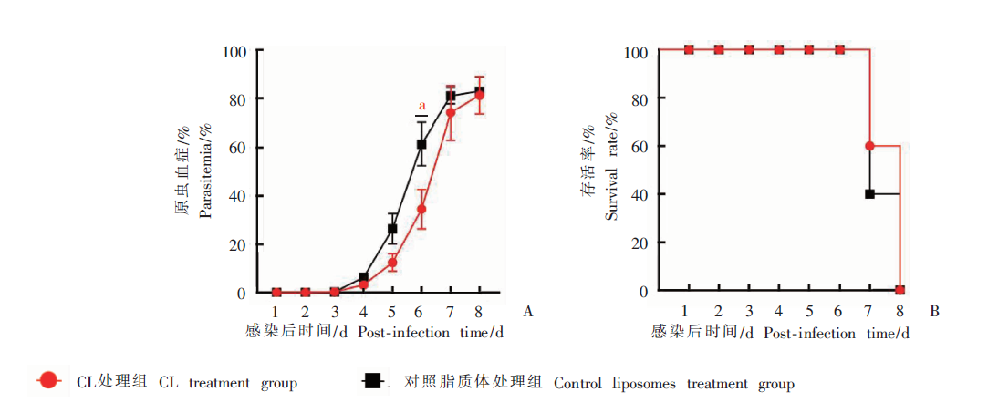
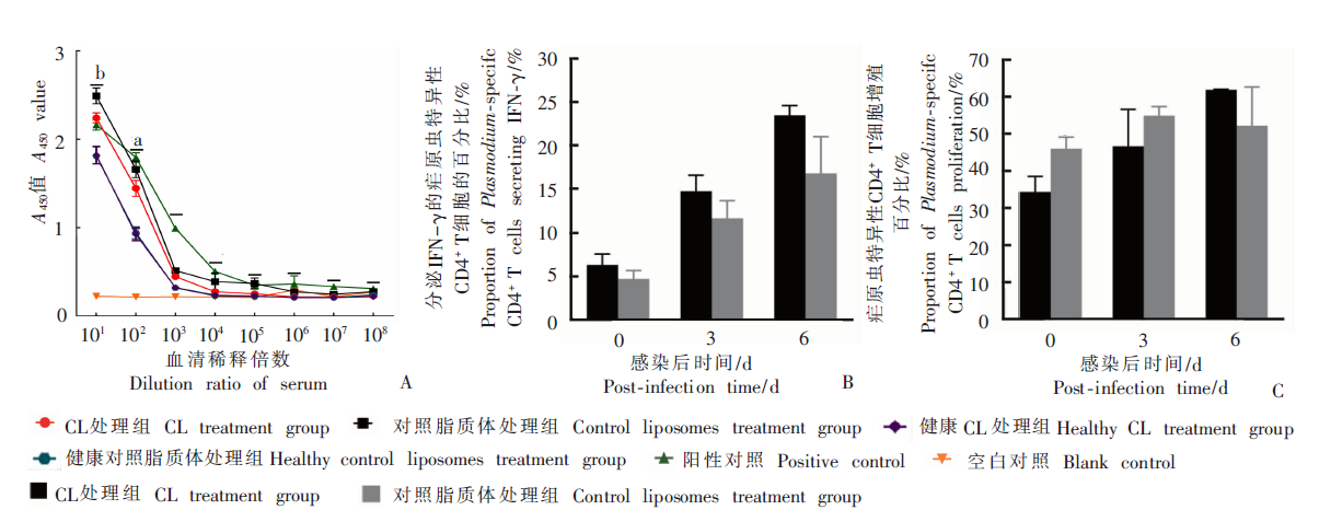
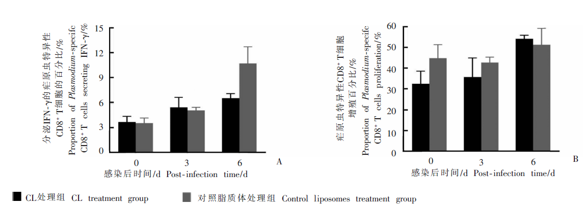
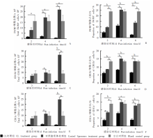
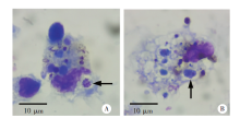
目的 探究高剂量氯磷酸脂质体(CL)处理对小鼠体内约氏疟原虫17XL(Py17XL)生长的影响。 方法 取61只雌性BALB/c小鼠,随机分成5组:高剂量CL处理组(简称CL处理组,23只)和对照脂质体处理组(29只)小鼠分别于感染Py17XL前1 d和感染后2、5 d,尾静脉注射高剂量CL(5 μg/μl 200 μl)和等量对照脂质体;健康CL处理组(3只)和健康对照脂质体处理组(3只)小鼠在相同时间尾静脉注射高剂量CL(5 μg/μl 200 μl)和等量对照脂质体;空白对照组(3只)小鼠不作任何处理。于感染后每天采集CL处理组和对照脂质体处理组小鼠(各5只)尾静脉血,涂片后显微镜下观察原虫血症,并统计小鼠存活情况。感染后0、3、6 d,取CL处理组和对照脂质体处理组小鼠(每次3只)脾脏,分离脾淋巴细胞,采用流式细胞术检测两组小鼠疟原虫特异性CD4+ T细胞和CD8+ T细胞的增殖能力和γ干扰素(IFN-γ)分泌水平,ELISA检测感染后6 d的小鼠血清抗疟原虫IgG抗体水平。感染后2、4、6 d,取CL处理组、对照脂质体处理组、空白对照组小鼠(每次3只)脾脏,分离脾淋巴细胞,采用流式细胞术检测CL耗竭的细胞种类,吉氏染色后显微镜下观察对照脂质体处理组小鼠(每次2只)脾脏树突状细胞的疟原虫感染情况。 结果 CL处理组和对照脂质体处理组小鼠均在感染后7 d出现死亡(CL处理组2只,对照脂质体处理组3只),且感染后8 d全部死亡,差异无统计学意义(χ2 = 0.360,P > 0.05)。感染Py17XL后,CL处理组小鼠的原虫血症均低于对照脂质体处理组小鼠,其中感染后6 d,CL处理组小鼠原虫血症为(34.537 ± 8.165)%,与对照脂质体处理组小鼠的(61.303 ± 8.799)%差异有统计学意义(F = 1.821,P < 0.05)。感染后2、4、6 d,CL处理组小鼠脾脏中的单核细胞(CD11b+)占比分别为(6.240 ± 0.605)%、(8.277 ± 0.411)%、(6.573 ± 0.246)%,树突状细胞(CD11c +)占比分别为(3.700 ± 0.599)%、(8.003 ± 0.655)%、(3.920 ± 0.534)%,巨噬细胞(F4/80 +)占比分别为(4.830 ± 0.695)%、(11.007 ± 1.121)%、(2.743 ± 0.395)%,均低于对应的对照脂质体处理组,且差异有统计学意义(F = 5.945、2.075、7.091,P < 0.05)。感染后3、6 d,CL处理组与对照脂质体处理组小鼠疟原虫特异性CD4 + T细胞和CD8+ T细胞的增殖能力和IFN-γ分泌水平差异均无统计学意义(P > 0.05)。感染后6 d,CL处理组小鼠稀释10倍的血清抗疟原虫IgG抗体水平为2.241 ± 0.056,低于对照脂质体处理组的2.490 ± 0.090,差异有统计学意义(F = 27.66,P < 0.05)。镜检结果显示,感染后2、4 d,对照脂质体处理组小鼠的CD11c+细胞内并未发现疟原虫,绝大多数感染后6 d的CD11c+细胞内发现了大量的疟原虫。 结论 高剂量CL耗竭巨噬细胞、树突状细胞和单核细胞后,并非通过调节小鼠抗红内期疟原虫的固有免疫和适应性免疫应答参与抑制Py17XL的增殖和发育。

关键词: 约氏疟原虫, 氯磷酸脂质体, 适应性免疫, 固有免疫, 树突状细胞

Abstract:

Objective To investigate the effect and mechanism of high-dose clodronate liposomes (CL) treatment on the growth of Plasmodium yoelii 17XL (Py17XL) in mice. Methods Sixty one female BALB/c mice were randomly assigned to five groups: high dose CL treatment group (CL treatment group, 23 mice) and control liposome treatment group (29 mice), healthy CL treatment group (3 mice) and healthy control liposome treatment group (3 mice), and blank control group (3 mice). The CL treatment group mice (23) were injected via tail vein with high-dose CL (5 μg/μl 200 μl) on d1 prior to infection and on d2 and d5 post-infection of Py17XL respectively; the control liposome treatment group mice (29) were injected intravenously with the same volume of control liposome at the respective time points. The healthy CL treatment group mice (3) and the healthy control liposome treatment group mice (3) were given intravenously with high-dose CL (5 μg/μl 200 μl) or the same amounts of control liposomes at the corresponding time points. The blank control group mice (3) were not treated. Tail vein blood samples from the mice of CL treatment group and control liposome treatment group(5 mice each) were collected daily post-infection, of which the smears were examined microscopically for estimating parasitemia and survival rate. Spleen samples were collected from the two groups on d0, d3 and d6 post-infection, and then the splenic lymphocytes were isolated to detect the proliferation of Plasmodium-specific CD4 + T cell and CD8+ T cells and the secretion of interferon γ (IFN-γ) using flow cytometry. The mice serum IgG antibodies against Plasmodium were determined by enzyme-linked immunosorbent assay (ELISA) on d6 post-infection. On d2, d4 and d6 post-infection, spleen lymphocytes of the CL treatment group, control liposome treatment group and blank control group (3 mice each time) were isolated, and the cell types depleted by CL were determined by flow cytometry. Plasmodium infection in the spleen dendritic cells of the control liposome treatment group (2 mice each time) was microscopically observed by Giemsa staining. Results In both the CL treatment group and control liposome treatment group, mice death occurred on d7 post-infection (2 in CL treatment group, 3 in control liposome treatment group), and all mice died on d8 post-infection, with no significant difference between the groups (χ 2 = 0.360, P > 0.05). After infection, the parasitemia in the CL treatment group was apparently lower than that in control liposome treatment group. On d6 post-infection, the parasitemia in the CL treatment group (34.537 ± 8.165)%, was significantly different from that in the control liposome treatment group (61.303 ± 8.799)% (F = 1.821, P < 0.05). On d2, d4 and d6 post-infection, the spleen cell responses in the CL treatment group were assayed, showing that the proportions of CD11b + monocytes in the spleen were (6.240 ± 0.605)%, (8.277 ± 0.411)%, (6.573 ± 0.246)%, and that of CD11c + dendritic cells in the spleen were (3.700 ± 0.599)%, (8.003 ± 0.655)%, (3.920 ± 0.534)%, and that of F4/80 + macrophages in the spleen were (4.830 ± 0.695)%, (11.007 ± 1.121)%, (2.743 ± 0.395)%, respectively, which were significantly lower than those in the control liposome treatment group (F = 5.945, 2.075, 7.091, P < 0.05). On d3 and d6 post-infection, no statistical differences were found in proliferation of Plasmodium-specific CD4 + T cells and CD8+ T cells, and secretion of IFN-γ between the CL treatment group and control liposome treatment group (P > 0.05). On d6 post-infection, the 10-fold-diluted serum IgG antibody level against Plasmodium in the CL treatment group (2.241 ± 0.056) was significantly lower than that of the control liposome treatment group (2.490 ± 0.090) (F = 27.66, P < 0.05). Microscopic examination indicated that no Plasmodium was observed by Giemsa staining in the CD11c+ cells of the control liposome treatment group on d2 and d4 post-infection, however, a large number of Plasmodium was found in almost all CD11c + cells on d6 post-infection. Conclusion After the macrophages, dendritic cells and monocytes were drained by high-dose CL intervention, regulation via innate immunity and adaptive immunity response against erythrocytic stage of Plasmodium in the mice may not involve in the suppression of Py17XL proliferation and growth.

Key words: Plasmodium yoelii, Clodronate liposomes, Adaptive immunity, Innate immunity, Dendritic cell

中图分类号: