中国寄生虫学与寄生虫病杂志 ›› 2018, Vol. 36 ›› Issue (6): 673-675.

• 研究简报 • 上一篇    下一篇

宁海县首例牛带绦虫感染病例的诊治分析

俞谊江1,*(), 郑静静1, 王斌1, 任丛汉1, 顾敏霞1, 徐海红2   

  1. 1 宁海县疾病预防控制中心,宁海315600
    2 宁海县前童镇中心卫生院,前童315636
  • 收稿日期:2018-06-13 出版日期:2018-12-30 发布日期:2019-01-08
  • 通讯作者: 俞谊江

A case report of Taenia saginata infection in Ninghai County: diagnosis and treatment

Yi-jiang YU1,*(), Jing-jing ZHENG1, Bin WANG1, Cong-han REN1, Min-xia GU1, Hai-hong XU2   

  1. 1 Ninghai Center for Disease Control and Prevention, Ninghai, 315600, China
    2 Health center of the Qiantong Town, Ninghai, 315636, China
  • Received:2018-06-13 Online:2018-12-30 Published:2019-01-08
  • Contact: Yi-jiang YU

摘要:

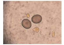
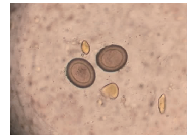
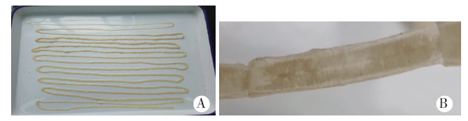
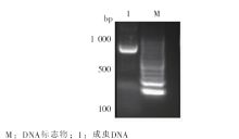
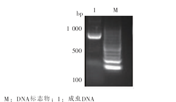
诊治分析宁海县首例带绦虫感染病例,并鉴定感染虫种。对患者开展流行病学调查,收集患者主要临床表现、生活习惯等资料。采集患者粪样,制备生理盐水涂片,观察虫卵形态。使用槟榔南瓜子进行驱虫治疗,对患者排出的虫体进行形态学观察。利用PCR技术扩增虫体DNA的线粒体细胞色素c氧化酶1(cox1)基因片段并进行序列分析。流行病学调查结果显示,患者无外出旅行史和生食史,仅2017年9月初曾食过未完全煮熟的牛肉。于2017年11月10日出现腹泻、易饥饿现象;11月26日,肛周首次排出长约30 cm、白色、扁平状、类似面条样的节片;12月4日,肛周再次排出长约2 cm的多节节片,遂至宁海县中医院诊治,诊断为“疑似带绦虫病”。粪样镜检结果显示,可见虫卵,内含六钩蚴,未见卵膜。对排出的成虫进行形态学鉴定:虫体前端细长,向后逐渐变粗变宽变厚,由颈节和链体节片连接而成,未发现头节;孕节子宫分支整齐,每侧约24~28支,总长5.32 m,疑似牛带绦虫。cox1基因经PCR扩增后,可获得长度为832 bp的目的条带。测序后的BLAST比对分析结果显示,目的条带与牛带绦虫(Taenia saginata,GenBank登录号为AB107239.1)cox1基因序列的同源性为99%,与亚洲带绦虫(Taenia asiatica,GenBank登录号为AB107235.1)和猪带绦虫(Taenia solium,GenBank登录号为AB066485.1)cox1基因序列的同源性分别为96%和88%。综合患者的流行病学调查、虫体形态学观察、cox1基因序列比对的结果,确诊该患者属于食用未煮熟牛肉引起的牛带绦虫感染。

关键词: 牛带绦虫, 诊断, 线粒体细胞色素c氧化酶1基因

Abstract:

Epidemiological information on the first case of taenia infection in Ninghai County was collected and analyzed. Eggs in the patient feces were founded using saline smear method. The patient was dewormed with areca and pumpkin seeds and the expelled worm was collected for morphological identification. The cytochrome c oxidase 1(cox1) gene was amplified from the worm using PCR. The epidemiological results showed that the patient had no history of travelling out of the living area, but once experienced eating undercooked beef in a hot pot in September, 2017. The patient started to have manifestation of diarrheal and hunger on November 10, 2017. A white, flat and noodle-like segment of worm with 30 cm in length was first observed in the feces 2-weeks later. One more segment with 2 cm long was then seen in feces. The patient went to see a doctor and was diagnosed as taeniasis. The eggs were observed in the fecal sample with an oncosphere inside without membrane. The expelled worm was total 5.32 m in length, with a slim and long anterior part and gradually wide body connected with segments. The scolex was not observed. The gravid proglottids contained 24-28 uterus branches on each side. The morphological examination showed consistency with that of Taenia saginata. A DNA fragment with 832 bp of cox1 were amplified by PCR and the sequence results showed 99%, 96% and 88% identity to cox1 of Taenia saginata, Taenia asiatica and Taenia solium respectively (GenBank accession number: AB107239.1, AB107235.1, AB066485.1). The results confirm that this is a case of Taenia saginata infection.

Key words: Taenia saginata, Diagnosis, Mitochondrial cytochrome c oxidase 1 gene

中图分类号: