中国寄生虫学与寄生虫病杂志 ›› 2018, Vol. 36 ›› Issue (6): 579-585.

• 论著 • 上一篇    下一篇

日本血吸虫感染小鼠肝脏和脾脏淋巴细胞及其表面程序性死亡配体1表达动态变化的研究

王晓玲, 胡媛*(), 徐馀信, 刘华, 曹建平   

  1. 中国疾病预防控制中心寄生虫病预防控制所,国家热带病研究中心,卫生部寄生虫病原与媒介生物学重点实验室,科技部国家级热带病国际联合研究中心,世界卫生组织热带病合作中心,上海 200025
  • 收稿日期:2018-07-05 出版日期:2018-12-30 发布日期:2019-01-08
  • 通讯作者: 胡媛
  • 基金资助:
    国家自然科学基金(No. 81772225);国家科技重大专项(No. 2018ZX10102001);上海市第四轮公共卫生三年行动计划(No. 15GWZK0101)

Dynamic changes of lymphocyte differentiation and programmed cell death protein ligand 1 expression in mice infected with Schistosoma japonicum

Xiao-ling WANG, Yuan HU*(), Yu-xin XU, Hua LIU, Jian-ping CAO   

  1. National Institute of Parasitic Diseases, Chinese Center for Disease Control and Prevention
    Chinese Center for Tropical Diseases Research
    Key Laboratory of Parasite and Vector Biology, Ministry of Health
    National Center for International Research on Tropical Diseases, Ministry of Science and Technology
    WHO Collaborating Centre for Tropical Diseases, Shanghai 200025, China
  • Received:2018-07-05 Online:2018-12-30 Published:2019-01-08
  • Contact: Yuan HU
  • Supported by:
    Supported by the National Natural Science Foundation of China (No. 81772225),National Science and Technology Major Project (No. 2018ZX10102001) and the Fourth Round of Three-Year Public Health Action Plan of Shanghai, China (No. 15GWZK0101)

摘要:

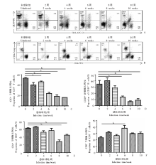
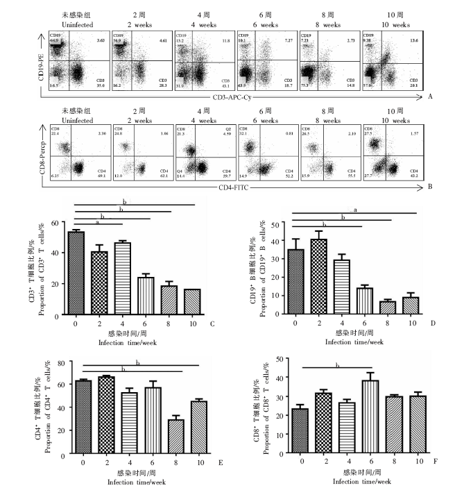
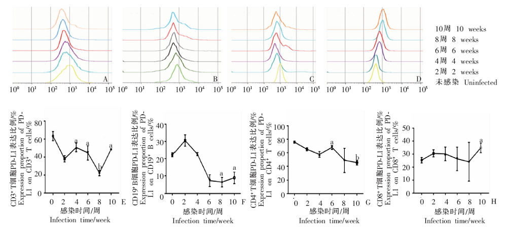
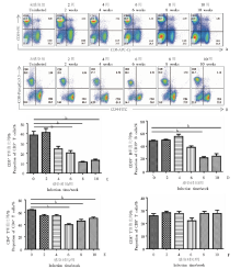
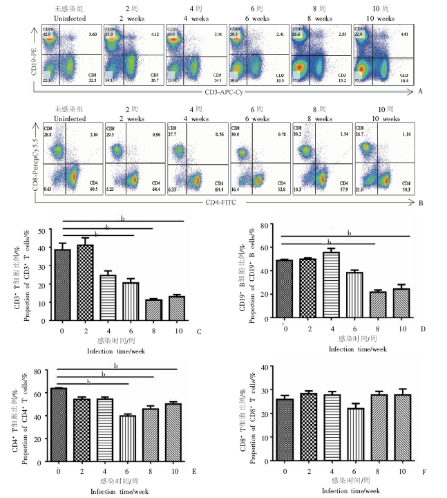
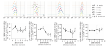
目的 初步研究日本血吸虫感染对小鼠肝脏和脾脏淋巴细胞数量及其表面程序性死亡配体1(PD-L1)功能的影响。方法 30只BALB/c小鼠随机分为感染组和未感染组,每组15只,感染组小鼠经腹部皮肤感染日本血吸虫尾蚴(20 ± 2)条/鼠。感染后2、4、6、8和10周,分别随机取两组小鼠各3只,颈椎脱臼处死小鼠,分离小鼠肝脏和脾脏的淋巴细胞,采用流式细胞术检测CD3+ T细胞(CD3+CD19-)、CD19+ B细胞(CD3-CD19+)、CD4+ T细胞(CD3+CD19-CD4+CD8-)和CD8+T细胞(CD3+CD19-CD4-CD8+)淋巴细胞比例的动态变化,以及这些细胞群上的PD-L1表达变化。组间差异比较采用ANOVA检验。结果 感染日本血吸虫后4~8周,小鼠肝脏和脾脏中的CD3+ T细胞、CD19+ B细胞和CD4+ T细胞比例均低于未感染组(P < 0.05或0.01,P > 0.05)。感染日本血吸虫后6周的小鼠肝脏中CD8+ T细胞比例增加[(38.03 ± 7.41)%],与未感染组[(23.37 ± 3.98)%]比较,差异有统计学意义(P < 0.01);但脾脏中无明显变化。在感染日本血吸虫后6周,小鼠肝脏中CD19+ B细胞上PD-L1的表达被显著抑制[(7.25 ± 3.47)%],与未感染组[(22.77 ± 8.90)%]比较,差异有统计学意义(P < 0.01);脾脏相对肝脏较晚,感染后8周CD19+ B细胞上PD-L1的表达被抑制[(22.37±4.01)%],与未感染组[(51.97 ± 1.62)%]比较,差异有统计学意义(P < 0.01),且此后在肝脏和脾脏中保持低水平表达。感染后2~10周,肝脏中的CD3+ T和CD4+ T细胞表面PD-L1的表达低于未感染组(P <0.05或0.01,P > 0.05),脾脏中的CD3+T和CD4+T细胞表面PD-L1的表达分别于感染后4~8周和6~10周低于未感染组(P <0.05)。肝脏和脾脏中CD8+ T细胞上PD-L1的表达在感染早期无变化,感染后10周比例增加,为(34.80 ± 3.68)%、(31.90 ± 2.53)%,与未感染组[(25.5 ± 0.80)%、(29.91 ± 3.55)%]比较,差异有统计学意义(P < 0.01)。结论 日本血吸虫感染BALB/c小鼠后,肝脏和脾脏中CD19+ B、CD3+ T和CD4+ T淋巴细胞亚群的比例显著降低,但对CD8+ T细胞比例影响不明显。感染对PD-L1在上述淋巴细胞亚群的表达也有相同的变化,在CD19+ B、CD3+ T和 CD4+ T细胞上表达受抑制,在CD8+ T细胞上的表达影响较小,且肝脏局部淋巴细胞的变化早于脾脏。

关键词: 日本血吸虫, 小鼠, 淋巴细胞, 流式细胞术, 程序性死亡配体1

Abstract:

Objective To evaluate the effect of Schistosoma japonicum infection on the lymphocyte differentiation and the programmed cell death protein ligand 1 (PD-L1) expression in infected mice. Methods Thirty BALB/c mice were randomly divided into infected group and uninfected groups. Mice in infected group were each infected with 20 ± 2 cercariae through abdominal skin. Three mice from each group were euthanized before infection and 2, 4, 6, 8, 10 weeks after infection and the lymphocytes were isolated from livers and spleens. The dynamic changes of the lymphocyte proportions for CD3+ T cells (CD3+CD19-), CD19+ B cells (CD3-CD19+), CD4+ T cells (CD3+CD19-CD4+CD8-) and CD8+ T cells(CD3+CD19-CD4-CD8+) and PD-L1 expression on these subset cells were detected by flow cytometry. The differences between groups were statistically analyzed by ANOVA test. Results Both in liver and spleen, the lymphocyte populations of CD3+ T cells, CD19+ B cells and CD4+ T subsets, decreased significantly in mice infected with S. japonicum for 4-8 weeks compared to the populations before infection (P < 0.05, 0.01 or P > 0.05). However, in livers of mice infected with S. japonicum, the population of CD8+ T cell subset at 6 week [(38.03 ± 7.41)%] after infection was significantly higher than uninfected group [(23.37 ± 3.98)%](P < 0.01), but no significant change in spleen. More interestingly, the expression of PD-L1 on CD19+ B cells at 6 week [(7.25 ± 3.47)%] after infection with S. japonicum was significantly inhibited compared with the uninfected group [(22.77 ± 8.90)%] (P < 0.01) while the changes in spleen was later than that in liver, at 8 week[(22.37±4.01)%] after infection was significantly inhibited compared with the uninfected group[(51.97 ± 1.62)%] (P < 0.01) and remained at low level thereafter, in both liver and spleen. The expression of PD-L1 on CD3+ T and CD4+ T cells in liver during 2 to 10 weeks (P < 0.05, 0.01 or P > 0.05) and in spleen during 4 to 8 weeks after infection was lower than that of the uninfected group (P <0.05). The expression of PD-L1 on CD8+ T cells in both liver and spleen was not changed at the early infection stage, but increased significantly at 10 weeks [(34.80 ± 3.68)%, (31.90 ± 2.53)%] after infection compared to the level before infection [(25.5 ± 0.80)%, (29.91 ± 3.55)%] (P < 0.01). Conclusion The lymphocyte proportion of CD19+ B, CD3+ T and CD4+ T cells were induced to decrease significantly in livers and spleens of mice infected with S. japonicum, however, the change in CD8+ T cell proportion was not significant in comparison to the level before infection. Similar trend was observed in PD-L1 expression on these lymphocyte subsets with significant decrease on CD19+ B, CD3+ T and CD4+ T lymphocytes, but significant increase on CD8+ T cells at later infection stage. These changes of lymphocytes in liver occur earlier than that in spleen.

Key words: Schistosoma japonicum, BALB/c mice, Lymphocyte, Flow cytometry, PD-L1

中图分类号: