中国寄生虫学与寄生虫病杂志 ›› 2017, Vol. 35 ›› Issue (3): 224-229.

• 论著 • 上一篇    下一篇

库普弗细胞在小鼠日本血吸虫肝病发展过程中的表型变化

孙悦1,2, 郑葵阳1,*(), 何兴2, 雷南行2, 颜超1, 汤仁仙1   

  1. 1徐州医科大学病原生物学与免疫学教研室,江苏省免疫与代谢重点实验室,徐州 221004
    2第二军医大学热带传染病学教研室,上海 200433
  • 收稿日期:2016-12-22 出版日期:2017-03-30 发布日期:2017-09-07
  • 通讯作者: 郑葵阳

The phenotype changes of Kupffer cells in the progression of hepatic schistosomiasis japonica

Yue SUN1,2, Kui-yang ZHENG1,*(), Xing HE2, Nan-hang LEI2, Chao YAN1, Ren-xian TANG1   

  1. 1 Department of Pathogen Biology and Immunity, Xuzhou Medical University; Jiangsu Province Key Laboratory of Immunometabolism, Xuzhou 221000, China
    2 Department of Tropical Infectious Diseases, The Second Military Medical University, Shanghai 200433, China
  • Received:2016-12-22 Online:2017-03-30 Published:2017-09-07
  • Contact: Kui-yang ZHENG

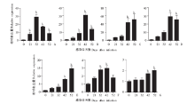
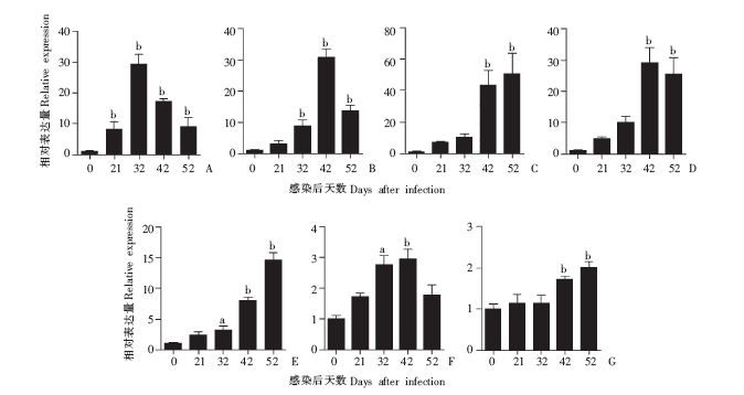
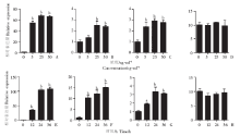
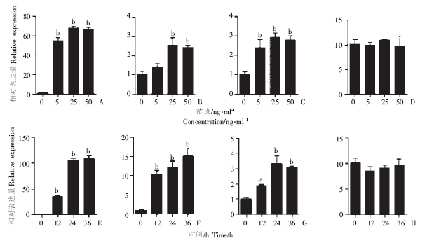
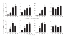
摘要: 目的 研究库普弗细胞(Kupffer cell)在小鼠日本血吸虫(Schistosoma japonicum)肝病发展过程中的表型变化。 方法 将20只6周龄的BALB/c雄性小鼠经腹部皮肤感染16条日本血吸虫尾蚴,在感染后0、21、32、42、52 d分别处死小鼠,取肝脏组织,HE染色和Masson三色染色观察肝脏病理变化,实时定量PCR(qPCR)检测肝脏Th1型细胞因子γ干扰素(interferon-γ,IFN-γ)、α肿瘤坏死因子(tumor necrosis factor-α,TNF-α)、Th2型细胞因子白细胞介素-4(interleukin-4,IL-4)、IL-13、IL-10)以及库普弗细胞的M1型巨噬细胞分化标志物诱导型一氧化氮合酶(inducible nitric oxide synthetase,iNOS)、白细胞分化抗原16(cluster of differentiation 16,CD16)、IL-6和M2型巨噬细胞分化标志物精氨酸酶-1(arginase 1,Arg-1)、CD206、IL-10的表达。体外培养库普弗细胞系,分别给予0、5、25、50 ng/ml IFN-γ或IL-4刺激12 h,或者给予25 ng/ml IFN-γ或IL-4分别刺激0、12、24、36 h后,qPCR检测库普弗细胞M1型、M2型巨噬细胞分化标志物。 结果 HE染色和Masson三色染色结果显示,小鼠感染后32 d肝组织中开始有虫卵沉积,42 d有明显的肉芽肿和纤维化病变。qPCR结果显示,与感染后0 d相比,IFN-γ在32 d表达水平最高,相对表达量为29.243 ± 3.245,52 d时迅速下降,为8.923 ± 3.002。IL-4在42 d最高,为25.521 ± 4.957。IL-13在52 d最高,为50.793 ± 9.631(均P < 0.05)。iNOS在42 d表达水平上升,相对表达量为2.950 ± 0.321,在52 d下降,为1.783 ± 0.319。Arg-1在52 d最高,为2.003 ± 0.152(均P < 0.05)。0、5、25、50 ng/mlIFN-γ刺激库普弗细胞12 h后,iNOS、IL-6和CD16的相对表达量分别为54.690~68.577、1.887~2.427、2.417~2.787(均P < 0.05)。25 ng/ml IFN-γ刺激0、12、24、36 h后,iNOS、IL-6和CD16的相对表达量分别为34.810~109.210、10.327~15.143、1.887~3.317(均P < 0.05),而Arg-1与对照组相比无明显变化。0、5、25、50 ng/ml IL-4刺激库普弗细胞12 h后,Arg-1、IL-10和CD206的相对表达量分别为9.153~24.253、1.923~3.687和37.770~72.133(均P < 0.05);25 ng/mlIL-4刺激0、12、24、36 h后Arg-1、IL-10和CD206的相对表达量分别为3.563~12.613、1.637~2.673和19.732~71.943(均P < 0.05),而iNOS无明显变化。 结论 在日本血吸虫感染早期,库普弗细胞以M1型为主;而在感染中晚期,库普弗细胞以M2型为主,表明库普弗细胞转变与肝脏免疫微环境密切相关.

关键词: 血吸虫病, 肉芽肿, 纤维化, 库普弗细胞

Abstract: Objective To observe the phenotype changes of Kupffer cells in the progression of hepatic schistosomiasis japonica. Methods Twenty male BALB/c mice at 6 weeks of age were infected with 16 cercariae of Schistosoma japonicum via a percutaneous route, then sacrificed at 0, 21, 32, 42 and 52 days after infection to collect liver samples for pathological observation by HE staining and Masson’s triple staining, and qPCR was used to detect the expression of Th1- [interferon-γ (IFN-γ) and tumor necrosis factor-α (TNF-α)] and Th2-type [IL-4 interleukin-4 (IL-4), IL-13 and IL-10] cytokines, as well as M1- [inducible nitric oxide synthetase (iNOS), cluster of differentiation 16(CD16) and IL-6] and M2-type [arginase 1 (Arg-1), CD206 and IL-10] markers of macrophages differentiated from Kupffer cells. Meanwhile, the Kupffer cells were treated with IFN-γ or IL-4 in vitro (0, 5, 25 and 50 ng/ml for 12 h; 25 ng/ml for 0, 12, 24 and 36 h), and the expression of M1- and M2-type markers was detected again by qPCR. Results HE staining and Masson’s triple staining revealed that the deposition of parasite eggs began to appear in the liver tissue from day 32 post-infection, and the formation of egg granulomas and fibrosis were observed on day 42 post-infection. qPCR results showed that the expression of IFN-γ in the liver tissue peaked on day 32 (29.243 ± 3.245) post-infection, and decreased sharply to 8.923 ± 3.002 on day 52 (P < 0.05 vs. day 0). The expression of IL-4 peaked on day 42 (25.521 ± 4.957), and that of IL-13 on day 52 (50.793 ± 9.631) (both P < 0.05). The expression of iNOS increased to 2.950 ± 0.321 on day 42, followed by a decrease to 1.783 ± 0.319 on day 52. The highest expression of Arg-1 was found on day 52(2.003 ± 0.152; P < 0.05). After 12 h treatment with 5, 25, and 50 ng/ml IFN-γ, the relative expression levels of iNOS, IL-6 and CD16 in Kupffer cells were dramatically elevated to 54.690-68.577, 1.887-2.427 and 2.417-2.787, respectively (P < 0.05 vs. control group), while those after 25 ng/ml IFN-γ treatment for 12, 24 and 36 h were 34.810-109.210, 10.327-15.143, and 1.887-3.317, respectively. However, no significant changes were found for Arg-1. The relative expression levels of Arg-1, IL-10 and CD206 were elevated significantly(9.153-24.253, 1.923-3.687 and 37.770-72.133, respectively) after 12 h treatment with 5, 25 and 50 ng/ml IL-4(P < 0.05), while those after 25 ng/ml IL-4 treatment for 12, 24 and 36 h were 3.563-12.613, 1.637-2.673, and 19.732-71.943. No significant changes were found for iNOS. Conclusion The Kupffer cell phenotype switches from M1 type in the early phase of infection to M2 type in the later period of infection, indicating a close relationship between Kupffer cell differentiation and liver immune microenvironment.

Key words: Schistosomiasis, Granuloma, Fibrosis, Kupffer cell

中图分类号: