中国寄生虫学与寄生虫病杂志 ›› 2023, Vol. 41 ›› Issue (2): 198-201.doi: 10.12140/j.issn.1000-7423.2023.02.011

• 论著 • 上一篇    下一篇

肝片形吸虫囊蚴脱囊条件及脱囊蚴形态观察

方文(), 杨敬, 王海英, 陈绍荣, 刘榆华, 李天美*()   

  1. 大理州血吸虫病防治研究所,云南大理 671000
  • 收稿日期:2022-06-28 修回日期:2022-10-28 出版日期:2023-04-30 发布日期:2023-05-10
  • 通讯作者: 李天美
  • 作者简介:方文(1966-),女,本科,主任医师,从事寄生虫病防治研究。E-mail:xfsfangwen@126.com
  • 基金资助:
    云南省医学学科后备人才培养项目(H-2018073)

Excystment conditions and excysted metacercaria morphological observation of Fasciola hepatica

FANG Wen(), YANG Jing, WANG Haiying, CHEN Shaorong, LIU Yuhua, LI Tianmei*()   

  1. The Institute of Research and Control on Schistosomiasis in Dali Prefecture, Dali 671000, Yunnan Province, China
  • Received:2022-06-28 Revised:2022-10-28 Online:2023-04-30 Published:2023-05-10
  • Contact: LI Tianmei
  • Supported by:
    Yunnan Province Medical Discipline Reserve Talent Training Program(H-2018073)

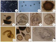
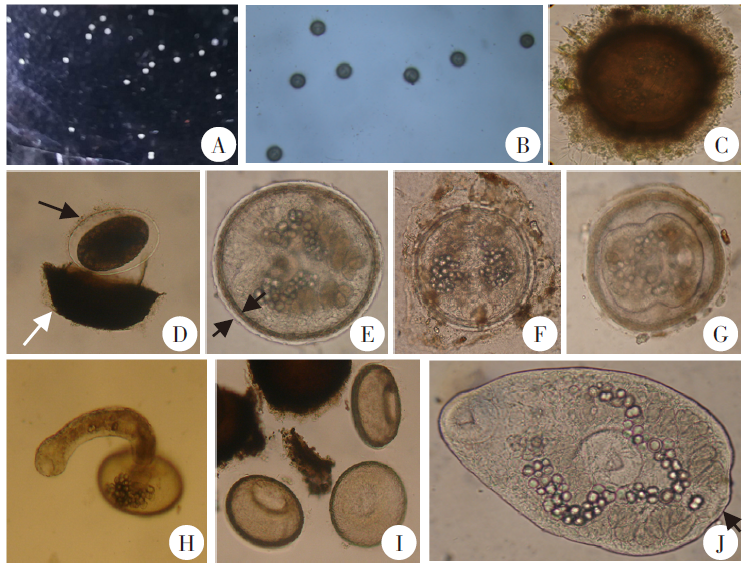
摘要: 目的 观察肝片形吸虫囊蚴脱囊所需条件及脱囊蚴的形态。 方法 收集牛源肝片形吸虫虫卵,28 ℃水浴10~11 d后,将孵化出的毛蚴感染小土蜗,感染后35~40 d收集囊蚴。将囊蚴分为3组,每组30个囊蚴,分别置于50%、75%和100%胆汁中,每组囊蚴再均分为2个小组,分别于37、39 ℃水浴,每隔30 min观察后尾蚴逸出情况,以选择最适脱囊温度。另设15个实验组,每组取10个囊蚴,囊蚴经人工胃液处理10 min、30 min、1 h、1.5 h和2 h后,分别加至50%、75%和100%胆汁中,用最适脱囊温度孵育6 h,每隔30 min观察脱囊蚴逸出情况,并镜下观察脱囊蚴形态。以蒸馏水、生理盐水、人工胃液以及50%、75%和100%胆汁直接处理为对照组。未孵育出脱囊蚴的囊蚴继续观察至24 h。结果 经不同温度孵育24 h后,39 ℃下脱囊蚴逸出数量最多,50%、75%、100%胆汁中分别逸出9、12和4条,因此最适水浴温度为39 ℃。不同实验组中,最早观察到脱囊蚴的为人工胃液处理2 h + 100%胆汁组,共耗时3 h(胆汁孵育1 h);逸出数量最多的为人工胃液处理1 h + 100%胆汁组,胆汁孵育6 h内共逸出8条脱囊蚴。6个对照组在观察时间内均未见脱囊蚴,但不同浓度胆汁处理24 h后可见已死亡的脱囊蚴。镜下可见,脱囊蚴呈长椭圆形,与尾蚴体部形态相似,为成虫的雏形,大小为(228.0~361.0)µm ×(114.0~209.0)µm,平均291.0 µm × 163.9 µm。口腹吸盘清晰可见;肠管呈黄褐色,自口吸盘后、咽及食管下,分两支盘曲重叠,充满虫体两侧直至尾部。 结论 肝片形吸虫囊蚴不会自行脱囊,脱囊过程需要胃肠消化液等因素的共同作用。脱囊蚴与尾蚴体部形态、大小相似。

关键词: 肝片形吸虫, 囊蚴, 脱囊, 脱囊蚴

Abstract: Objective To observe the required conditions of excystment and morphology of metacercariae of Fasciola hepatica. Methods Eggs of F. hepatica from cattle were collected and placed in 28 ℃ water bath for 10 or 11 days to hatch miracidia, which were used to infect Galba pervia for 35-40 days for collection of metacercariae. The metacercariae were assigned to 3 groups with 30 in each group. The metacercariae were placed in bovine bile at 50%, 75% and 100% dilution, respectively. Each group of metacercariae was futher divided into 3 groups and placed in water baths at 37 ℃ and 39 ℃ to observe emerging excysted worms every 30 min for ascertaining the optimal excystment temperature. In addition, 15 experimental groups of metacercariae were set up with 10 in each group to examine the excyst condition. After treatment with artificial gastric juice for 10 min, 30 min, 1 h, 1.5 h and 2 h, the cysts were transferred into 50%, 75% and 100% bovine bile, respectively, and incubated at the optimal temperature for 6 h to observe the exeyt process by inspection every 30 min. The morphology of excysted worms were examined microscopically. For control group, distilled water, normal saline, artificial gastric juice and 50%, 75% and 100% bovine bile were used alone, respectively. For those metacercarae that has not excysted, incubation will be continued in water bath for 24 h upon observation. Results After incubation at 37 ℃ and 39 ℃ separately for 24 h, highest yield of excysted worms was seen at 39 ℃, with 9, 12 and 4 excysted cercariae observed in 50%, 75% and 100% bile, respectively. It was indicated that the optimal incubation temperature for excystment in bile was 39 ℃. The excysted cercariae were earliest seen after incubation in artificial gastric juice for 2 h coupling with 100% bile for 1 h. The highest number of excysted cercariae was found in artificial gastric juice incubation for 1 h coupling with 100% bile 6 h, yieling 8 excysted cercariae. No excyst was observed in the 6 control groups during the observation period, but dead excysted worms were seen after 24 h in different concentrations of bile. Under microscope, the excysted cercariae were shaped in long oval, similar to the cercariae body in morphology, which become the juvenile form of adult worm. The size of excysted cercariae was (228.0-361.0) μm × (114.0-209.0) μm, with an average of 291.0 μm × 163.9 μm, having clearly visible oral and ventral suckers; the intestinal duct is yellowish brown in color, extending from the oral sucker, pharynx and esophagus to two branches in coiled overlapping, being full of both sides of worm body through to the tail. Conclusion F. hepatica metacercariae are unable to excyst spontaneously, which requires gastrointestinal digestive fluid factors to co-activate. The body morphology and size of excysted worms are similar to those of cercariae.

Key words: Fasciola hepatica, Metacercariae, Excystment, Decysticercosis

中图分类号: