中国寄生虫学与寄生虫病杂志 ›› 2022, Vol. 40 ›› Issue (3): 305-308.doi: 10.12140/j.issn.1000-7423.2022.03.021

• 研究简报 • 上一篇    下一篇

华支睾吸虫高迁移率族蛋白1同源蛋白的表达及其对小鼠巨噬细胞核转录因子-κB活化作用

王婷(), 杨庆利(), 冷静, 李宝莹, 申继清, 戴悦   

  1. 广西中医药大学,广西高发传染病中西医结合转化医学重点实验室,南宁 530200
  • 收稿日期:2021-09-09 修回日期:2021-12-21 出版日期:2022-06-30 发布日期:2022-07-06
  • 通讯作者: 杨庆利
  • 作者简介:王婷(1994-),女,硕士研究生,从事病原分子生物学研究。E-mail: 981693327@qq.com
  • 基金资助:
    广西自然科学基金(2020GXNSFAA238016);广西中医药大学博士科研启动基金(2019BS027)

Expression of the high mobility group box 1 homologous protein of Clonorchis sinensis and its effect on nuclear transcription factor-κB activation in mouse macrophages

WANG Ting(), YANG Qing-li(), LENG Jing, LI Bao-ying, SHEN Ji-qing, DAI Yue   

  1. Guangxi University of Chinese Medicine,Guangxi Key Laboratory of Translational Medicine for Treating High-Incidence Infectious Diseases with Integrative Medicine,Nanning 530200,China
  • Received:2021-09-09 Revised:2021-12-21 Online:2022-06-30 Published:2022-07-06
  • Contact: YANG Qing-li
  • Supported by:
    Natural Science Foundation of Guangxi(2020GXNSFAA238016);Doctoral Research Foundation of Guangxi University of Traditional Chinese Medicine(2019BS027)

摘要:

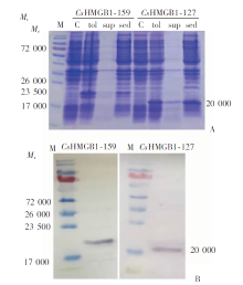
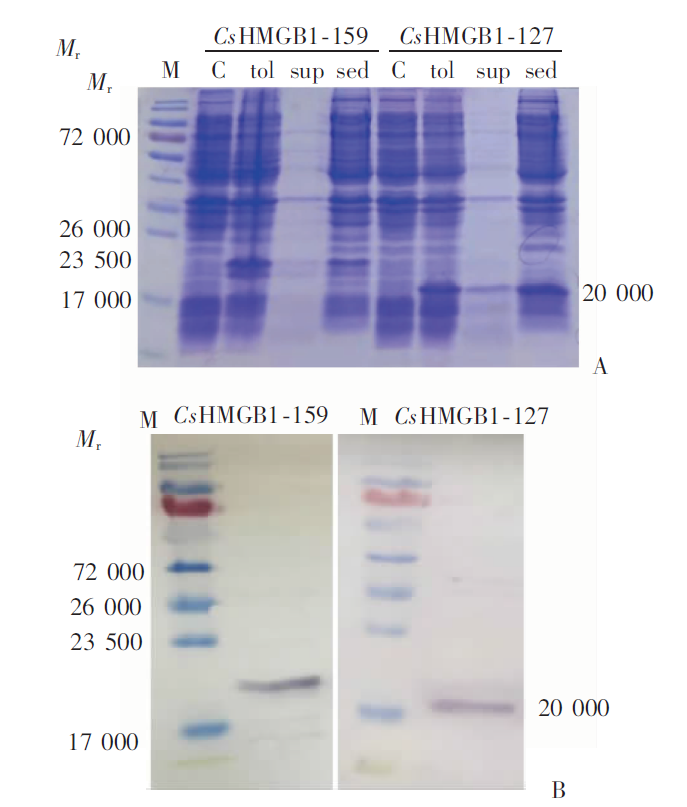
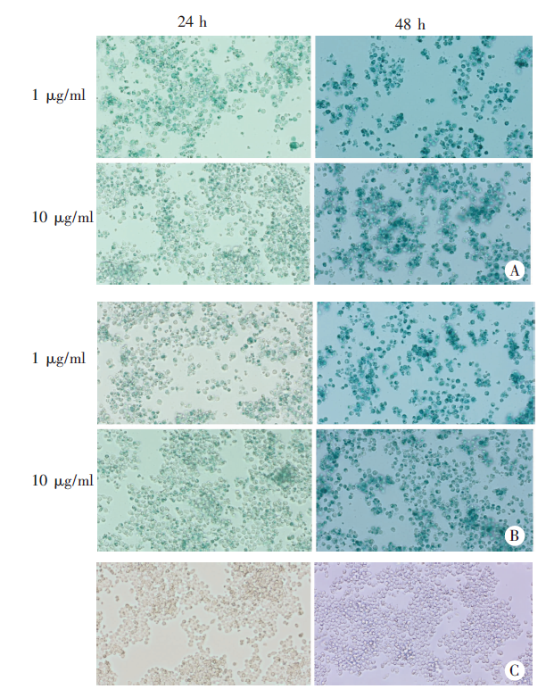
根据华支睾吸虫死亡细胞释放的高迁移率族蛋白1(CsHMGB1)同源分子编码序列构建pET-28a(+)表达质粒并转化至Rosetta(DE3)感受态细胞,异丙基-β-D-硫代半乳糖苷(IPTG)诱导CsHMGB1-His融合蛋白表达;Ni-TED琼脂糖树脂纯化蛋白后,蛋白质免疫印迹(Western blotting)分析鉴定His标签融合蛋白表达情况。用含不同浓度(1 μg/ml和10 μg/ml)CsHMGB1-159/127的培养液与小鼠巨噬细胞RAW264.7共培养,同时设立无刺激物的空白对照组,培养24 h和48 h。用pNiFty2-SEAP质粒和HEK-BlueTM培养基检测SEAP活性代表核转录因子-κB(NF-κB)活化,用酶标仪检测培养上清吸光度(A620值),并在显微镜下观察细胞内蓝色显色反应。结果显示,分别表达出编码159和127个氨基酸的CsHMGB1-159/127蛋白,相对分子质量(Mr)约为23 500和20 000。1和10 μg/ml CsHMGB1-159蛋白刺激小鼠巨噬细胞24 h后的A620值分别为0.66 ± 0.08和0.65 ± 0.03,均高于对照组(0.29 ± 0.02)(t = 11.1、28.5,P < 0.01),且胞浆均出现明显蓝色;48 h后的A620值分别达1.02 ± 0.08和1.07 ± 0.08,均高于对照组(0.62 ± 0.035)(t = 11.2、12.9,P < 0.01),且胞浆蓝色加深。用1和10 μg/ml CsHMGB1-127蛋白刺激24 h后的A620值分别为0.52 ± 0.08和0.56 ± 0.08,均高于对照组(t = 7.39、8.37,P < 0.01),胞浆也出现明显蓝色;48 h后的A620值分别达到1.10 ± 0.05和0.90 ± 0.10,均高于对照组(t = 20.30、6.26,P < 0.01),胞浆蓝色也明显加深。1和10 μg/ml CsHMGB1-159蛋白刺激24 h后的A620值高于相应浓度的CsHMGB1-127蛋白(t = 2.98、2.75,P < 0.05);48 h后两种蛋白的A620值的差异有统计学意义(t = -2.07,P < 0.01;t = 3.40,P < 0.05)。CsHMGB1-159比CsHMGB1-127蛋白在体外均能够刺激小鼠巨噬细胞NF-κB活化,CsHMGB1-159比CsHMGB1-127蛋白的作用更强。

关键词: 华支睾吸虫, 高迁移率族蛋白1, 病原相关分子模式, 核转录因子-κB

Abstract:

The PET-28a(+)expression plasmids were constructed according to the identified HMGB1 homologous gene sequences (CDSs)of Clonorchis sinensis and cloned into Rosetta (DE3) competent cells. Isopropyl-β-D-thiogalactoside (IPTG) was used to induce the expression of CsHMGB1-His fusion proteins. After purification by Ni-TED agarose resin, the expression of His-tagged fusion proteins was confirmed by Western blotting analysis. The mouse macrophage RAW264.7 cells were cultured with mediums supplemented with CsHMGB1-159/127 at a concentration of 1 μg/ml and 10 μg/ml, for 24 h and 48 h, respectively. The cells cultured without stimulants were used as the blank control. The SEAP activity, which indicates NF-κB activation, was detected by using pNiFty2-SEAP plasmid and HEK-BlueTM culture medium. The A620 value of culture supernatant was detected by microplate a reader, and the intracellular NF-κB activation was also observed under a microscope. Our results showed that the CsHMGB1-159/127 proteins encoding 159 and 127 amino acids were expressed, and the relative molecular weights (Mr) were approximately 23 500 and 20 000, respectively. The A620 values of supernatant from mouse macrophage stimulated with CsHMGB1-159 protein (1 and 10 μg/ml)for 24 hours were 0.66 ± 0.08 and 0.65 ± 0.03, respectively, which were higher than those in the control group (0.29 ± 0.02)(t = 11.1, 28.5; P < 0.01), and blue staining was observed in the cytoplasm. The A620 values after 48 h reached 1.02 ± 0.08 and 1.07 ± 0.08, respectively, which were higher than those in the control group (0.62 ± 0.035)(t = 11.2, 12.9; P < 0.01), and the blue staining in the cytoplasm was stronger. Similarly, the A620 values after 24 h stimulation with CsHMGB1-127 protein (1 and 10 μg/ml) were 0.52 ± 0.08 and 0.56 ± 0.08, respectively, which were higher than those in the control group(t =7.39, 8.37; P < 0.01), and the blue staining was observed in the cytoplasm. The A620 values after 48 h reached 1.10 ± 0.05 and 0.90 ± 0.10, respectively, which were also higher than those in the control group (t = 20.30, 6.26; P < 0.01), and stronger blue staining was also observed in the cytoplasm. The A620 values of CsHMGB1-159 protein (1 and 10 μg/ml) after 24 hours of stimulation were significantly higher than that of CsHMGB1-127 protein (t = 2.98, 2.75; P < 0.05). There were also significant differences in A620 value after 48 h stimulation (t = 3.40, P < 0.01). The HMGB1 homologous proteins of Clonorchis sinensis encoding 159 and 127 amino acids can stimulate mouse macrophage NF-κB activation in vitro. The CsHMGB1-159 has stronger stimulatory effect than CsHMGB1-127 protein.

Key words: Clonorchis sinensis, High mobility group box 1, Pathogen-associated molecular patterns, Nuclear transcription factor-κB

中图分类号: