| [1] |
Zhang MY,, Wu WP,, Guan YY, et al. Analysis on disease burden of hydatid disease in China[J]. Chin J Parasitol Parasit Dis, 2018, 36(1): 15-19, 25. (in Chinese)
|
|
( 张梦媛,, 伍卫平,, 官亚宜, 等. 我国棘球蚴病疾病负担分析[J]. 中国寄生虫学与寄生虫病杂志, 2018, 36(1): 15-19, 25.)
|
| [2] |
Wang TP,, Cao ZG. Current status of echinococcosis control in China and the existing challenges[J]. Chin J Parasitol Parasit Dis, 2018, 36(3): 291-296. (in Chinese)
|
|
( 汪天平,, 操治国. 中国棘球蚴病防控进展及其存在的问题[J]. 中国寄生虫学与寄生虫病杂志, 2018, 36(3): 291-296.)
|
| [3] |
Wei SH,, Wu WP,, Han S, et al. Analysis of the results of echinococcosis surveillance in China from 2016 to 2017[J]. J Pathog Biol, 2020, 15(8): 924-928. (in Chinese)
|
|
( 魏思慧,, 伍卫平,, 韩帅, 等. 2016—2017年全国棘球蚴病监测结果分析[J]. 中国病原生物学杂志, 2020, 15(8): 924-928.)
|
| [4] |
Shang JY,, Zhang GJ,, He W, et al. Taxonomy and molecular epidemiology of Echinococcus granulosus complex causing cystic echinococcosis[J]. Chin J Parasitol Parasit Dis, 2018, 36(2): 166-173, 177. (in Chinese)
|
|
( 尚婧晔,, 张光葭,, 何伟, 等. 细粒棘球蚴病病原分类学与分子流行病学研究进展[J]. 中国寄生虫学与寄生虫病杂志, 2018, 36(2): 166-173, 177.)
|
| [5] |
Chen J,, Wen H. Progress in diagnosis and treatment of hepatic echinococcosis[J]. J Southeast Univ Med Sci Ed, 2018, 37(5): 929-934. (in Chinese)
|
|
陈骏,, 温浩. 肝棘球蚴病的诊断与治疗进展[J]. 东南大学学报(医学版), 2018, 37(5): 929-934.)
|
| [6] |
Zhao SY,, Zhu HH,, Wang XQ, et al. Present situation and progress of comprehensive treatments for hepatic alveolar echinococcosis[J]. Chin J Schisto Control, 2019, 31(6): 676-678. (in Chinese)
|
|
( 赵顺云,, 朱海宏,, 王向前, 等. 肝多房棘球蚴病的综合治疗现状和进展[J]. 中国血吸虫病防治杂志, 2019, 31(6): 676-678.)
|
| [7] |
Chinese Congress of Hepatobiliary Surgeons. Expert consensus on diagnosis and treatment of hepatic hydatidosis(2015 Edition)[J]. Chin J Digest Surg, 2015, 14(4): 253-264. (in Chinese)
|
|
中国医师协会外科医师分会包虫病外科专业委员会. 肝两型包虫病诊断与治疗专家共识(2015版)[J]. 中华消化外科杂志, 2015, 14(4): 253-264.)
|
| [8] |
Sichuan Echinococcosis Diagnosis and Treatment Expert Group. Diagnosis and treatment of liver echinococcosis in Sichuan Province[J]. Chin J Bases Clin Gen Surg, 2017, 24(7): 798-803. (in Chinese)
|
|
( 四川省包虫病诊疗专家组. 四川省肝包虫病诊治规范[J]. 中国普外基础与临床杂志, 2017, 24(7): 798-803.)
|
| [9] |
Xiong YH,, Zheng B. The analytical research on patented technology of drugs for echinococcosis prevention and control in China[J]. Chin Health Stand Manag, 2021, 12(9): 5-9. (in Chinese)
|
|
( 熊彦红,, 郑彬. 中国棘球蚴病防治药物专利技术分析研究[J]. 中国卫生标准管理, 2021, 12(9): 5-9.)
|
| [10] |
Wang J,, Chen JY. Research progress of extracellular vesicles[J]. Chin J Tissue Eng Res, 2017, 21(4): 621-626. (in Chinese)
|
|
( 王琎,, 陈建英. 细胞外囊泡研究新进展[J]. 中国组织工程研究, 2017, 21(4): 621-626.)
|
| [11] |
Zhai JT,, Ma F. Advances in tumor cell-derived chemotherapeutic microparticles research[J]. Chin J Front Med Sci (Electron Version), 2020, 12(3): 27-30. (in Chinese)
|
|
翟婧彤,, 马飞. 肿瘤细胞来源的载药囊泡研究进展[J]. 中国医学前沿杂志(电子版), 2020, 12(3): 27-30.)
|
| [12] |
Yuan FM,, Li YM,, Wang ZH. Preserving extracellular vesicles for biomedical applications: consideration of storage stability before and after isolation[J]. Drug Deliv, 2021, 28(1): 1501-1509.
doi: 10.1080/10717544.2021.1951896
|
| [13] |
Chen Y. Research progress of extracellular vesicles[J]. Chin J Cell Biol, 2019, 41(2): 202-210. (in Chinese)
|
|
( 陈扬. 细胞外囊泡研究进展[J]. 中国细胞生物学学报, 2019, 41(2): 202-210.)
|
| [14] |
Meng WR,, He CS,, Hao YY, et al. Prospects and challenges of extracellular vesicle-based drug delivery system: considering cell source[J]. Drug Deliv, 2020, 27(1): 585-598.
doi: 10.1080/10717544.2020.1748758
|
| [15] |
Vader P,, Mol EA,, Pasterkamp G, et al. Extracellular vesicles for drug delivery[J]. Adv Drug Deliv Rev, 2016, 106(Pt A): 148-156.
|
| [16] |
Chen B,, Zhang Y,, Tang K. The invention relates to a storage method for tumor cell vesicle preparation[P]: China, CN109200029A. 2019-01-15. (in Chinese)
|
|
( 陈彬,, 张一,, 唐科. 一种肿瘤细胞囊泡制剂的贮存方法[P]: 中国, CN109200029A. 2019-01-15.)
|
| [17] |
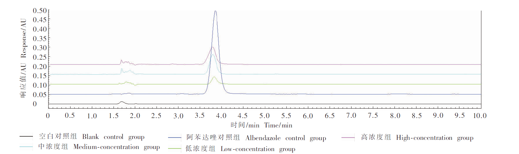
Zhao Y,, Zhang P,, Tang WB, et al. Determination of albendazole in albendazole tablets by high performance liquid chromatography[J]. Chin J Vet Drug, 2004, 38(5): 33-34, 37. (in Chinese)
|
|
( 赵英,, 张平,, 唐文标, 等. 高效液相色谱法测定兽用阿苯达唑片的含量[J]. 中国兽药杂志, 2004, 38(5): 33-34, 37.)
|
| [18] |
Zu YZ,, Tao DY,, Fang WS, et al. Status of drug treatment and prevention of echinococcosis[J]. J Clin Med Lit, 2017, 4(27): 5344, 5346. (in Chinese)
|
|
( 祖逸峥,, 陶栋义,, 方万胜, 等. 包虫病药物治疗与预防现状[J]. 临床医药文献电子杂志, 2017, 4(27): 5344, 5346.)
|
| [19] |
Pugholm LH,, Revenfeld ALS,, Søndergaard EKL, et al. Antibody-based assays for phenotyping of extracellular vesicles[J]. Biomed Res Int, 2015, 2015: 524817.
|
| [20] |
Li L,, Piontek K,, Ishida M, et al. Extracellular vesicles carry microRNA-195 to intrahepatic cholangiocarcinoma and improve survival in a rat model[J]. Hepatology, 2017, 65(2): 501-514.
doi: 10.1002/hep.28735
|
| [21] |
Zakharova L,, Svetlova M,, Fomina AF. T cell exosomes induce cholesterol accumulation in human monocytes via phosphatidy-lserine receptor[J]. J Cell Physiol, 2007, 212(1): 174-181.
pmid: 17299798
|
| [22] |
Chaput N,, Théry C. Exosomes: immune properties and potential clinical implementations[J]. Semin Immunopathol, 2011, 33(5): 419-440.
doi: 10.1007/s00281-010-0233-9
|
| [23] |
Ma JW,, Zhang Y,, Tang K, et al. Reversing drug resistance of soft tumor-repopulating cells by tumor cell-derived chemotherapeutic microparticles[J]. Cell Res, 2016, 26(6): 713-727.
doi: 10.1038/cr.2016.53
|
| [24] |
Gao YF,, Zhang H,, Zhou NN, et al. Methotrexate-loaded tumour-cell-derived microvesicles can relieve biliary obstruction in patients with extrahepatic cholangiocarcinoma[J]. Nat Biomed Eng, 2020, 4(7): 743-753.
doi: 10.1038/s41551-020-0583-0
|
| [25] |
Wang LZ,, Liu LK,, Liu J. Advances in isolation and purification techniques for exosomes[J]. Chemistry, 2021, 84(10): 1023-1030. (in Chinese)
|
|
( 王立志,, 刘路宽,, 刘晶. 外泌体分离与纯化技术研究进展[J]. 化学通报, 2021, 84(10): 1023-1030.)
|
| [26] |
Shan ZM,, Tao SC,, Hu CM, et al. Extraction, identification and proteomic analysis of exosomes derived from human umbilical cord mesenchymal stem cells[J]. Chin J Tissue Eng Res, 2022, 26(19): 3036-3042. (in Chinese)
|
|
( 单政铭,, 陶述春,, 胡春梅, 等. 人脐带间充质干细胞来源外泌体的提取、鉴定和蛋白组学分析[J]. 中国组织工程研究, 2022, 26(19): 3036-3042.)
|
| [27] |
Jäger R,, Zwacka RM. The enigmatic roles of caspases in tumor development[J]. Cancers, 2010, 2(4): 1952-1979.
doi: 10.3390/cancers2041952
|
| [28] |
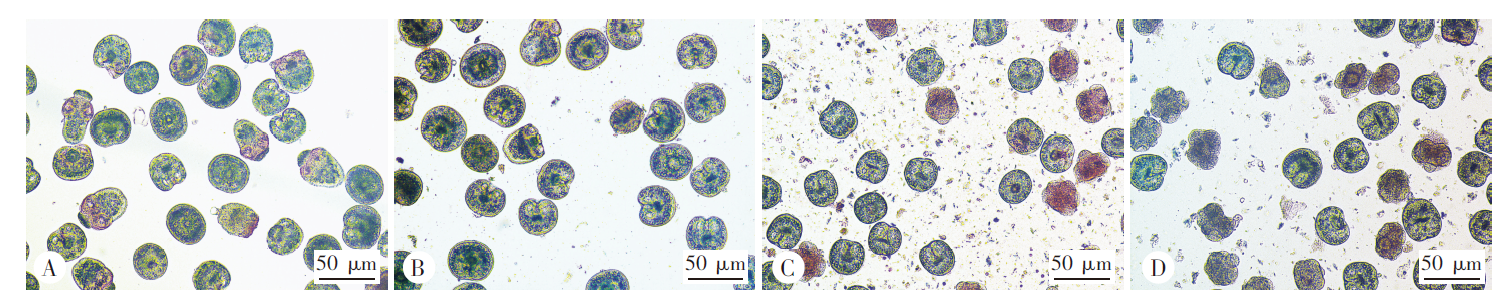
Kang JF,, Hu HH,, Chen R, et al. Observation on the apoptosis in protoscolex of hydatid cyst[J]. Chin J Zoonoses, 2010, 26(5): 433-435, 441. (in Chinese)
|
|
( 康金凤,, 胡汉华,, 陈蓉, 等. 棘球蚴原头节细胞凋亡的观察[J]. 中国人兽共患病学报, 2010, 26(5): 433-435, 441.)
|

 ), 周雪1, 刘程豪1, 姜慧娇1, 卜媛媛1, 陈雪玲2, 吴向未1,*(