中国寄生虫学与寄生虫病杂志 ›› 2021, Vol. 39 ›› Issue (6): 779-783.doi: 10.12140/j.issn.1000-7423.2021.06.008

• 论著 • 上一篇    下一篇

核因子-κB/髓样分化分子88在细粒棘球蚴病患者肝纤维化中的作用

马文梅1(), 桑伟1, 艾麦提·牙森2, 佐力克1, 付莉1, 苗娜1,*()   

  1. 1 新疆医科大学第一附属医院病理科,乌鲁木齐 830054
    2 新疆医科大学第一附属医院肝胆包虫病外科,乌鲁木齐 830054
  • 收稿日期:2021-06-28 修回日期:2021-09-02 出版日期:2021-12-30 发布日期:2021-12-15
  • 通讯作者: 苗娜
  • 作者简介:马文梅(1990-),女,硕士,初级技师,从事肝脏损伤与修复的分子机制研究。E-mail: marge10@126.com
  • 基金资助:
    省部共建中亚高发病成因与防治国家重点实验室开放课题资助项目(SKL-HIDCA-2018-30);省部共建中亚高发病成因与防治国家重点实验室开放课题资助项目(SKL-HIDCA-2020-28)

Roles of nuclear factor-κB/myeloid differentiation factor 88 in the liver fibrosis of cystic echinococcosis patients

MA Wen-mei1(), SANG Wei1, AIMAITI Ya-sen2, ZUO Li-ke1, FU Li1, MIAO Na1,*()   

  1. 1 Pathology Department, the First Affiliated Hospital of Xinjiang Medical University, Urumqi 830054, China
    2 Hepatobiliary and Echinococcosis Surgery Department, the First Affiliated Hospital of Xinjiang Medical University, Urumqi 830054, China
  • Received:2021-06-28 Revised:2021-09-02 Online:2021-12-30 Published:2021-12-15
  • Contact: MIAO Na
  • Supported by:
    State Key Laboratory of Pathogenesis, Prevention and Treatment of Central Asian High Incidence Diseases Fund(SKL-HIDCA-2018-30);State Key Laboratory of Pathogenesis, Prevention and Treatment of Central Asian High Incidence Diseases Fund(SKL-HIDCA-2020-28)

摘要:

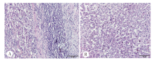
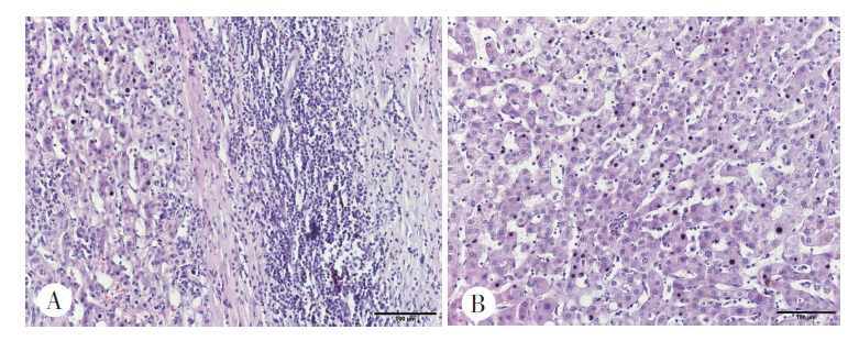
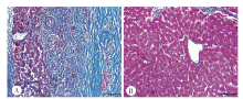
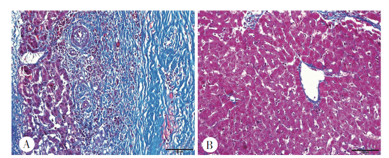
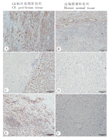
目的 探讨核因子-κB(nuclear factor-κB,NF-κB)/髓样分化分子88(myeloid differentiation factor 88,MyD88)在细粒棘球蚴病(cystic echinococcosis,CE)患者肝纤维化中的作用。 方法 收集CE患者手术切除的病灶肝组织,取距病灶近端(距病灶外囊壁0.5 cm内)和远端健康肝组织(距病灶外囊壁2 cm),HE染色观察肝组织病理变化。Masson染色观察胶原沉积情况。免疫组织化学染色和实时荧光定量PCR法(qRT-PCR)检测肝星状细胞活化标志物α-平滑肌肌动蛋白(α-smooth muscle actin,α-SMA)、NF-κB p65和MyD88在CE患者肝组织中的表达情况。两组均数比较采用独立样本t检验,采用Pearson相关系数法分析NF-κB p65、MyD88表达水平与患者肝组织胶原沉积阳性区域面积、α-SMA表达水平的相关性。 结果 共收集40份CE患者手术切除的肝组织病灶样品,其中CE1型8份、CE2型10份、CE3型7份、CE4型9份、CE5型6份。手术患者男性24例,女性16例,年龄20~45岁,平均(38.2 ± 12.9)岁。所取样品HE染色结果显示,CE病灶与近端肝组织间形成炎症微环境,呈现大量淋巴细胞浸润,伴不同程度的粒细胞增多。Masson染色结果显示,胶原主要沉积在病灶近端炎性细胞带、汇管区、胆管和血管周围,病灶近端肝组织的胶原沉积阳性区域面积为(20.29 ± 3.96)%,高于远端健康肝组织的(2.87 ± 1.74)%(t = 13.640,P < 0.01)。免疫组织化学染色结果显示,α-SMA、NF-κB p65及MyD88阳性细胞主要在CE患者病灶周围炎性细胞带中高表达,分别为(13.47 ± 3.47)%、(7.30 ± 1.40)%、(7.47 ± 1.86)%),在远端健康肝组织中仅少量表达,分别为(3.43 ± 0.56)%、(1.08 ± 0.29)%、(0.36 ± 0.05)%),二者差异有统计学意义(t = 5.682、18.530、5.087,P < 0.01)。qRT-PCR结果显示,在病灶近端肝组织中α-SMA、NF-κB p65和MyD88 mRNA的相对表达量分别为5.05 ± 0.42、3.71 ± 0.33、7.11 ± 0.50,远端健康肝组织分别为1.07 ± 0.01、1.02 ± 0.03、1.07 ± 0.02,二者差异具有统计学意义(t = 39.750、34.130、50.960,P < 0.01)。NF-κB p65、MyD88的表达水平与胶原沉积阳性区域面积、α-SMA的表达水平呈正相关关系(r = 0.98、0.97、0.98,P < 0.01)。 结论 NF-κB/MyD88在CE患者病灶近端肝组织的表达较远端健康肝组织明显升高,并与肝纤维化程度呈正相关。

关键词: 肝细粒棘球蚴病, 核因子-κB, 髓样分化分子88, 肝纤维化

Abstract:

Objective To investigate roles of nuclear factor-κB/myeloid differentiation factor 88 (NF-κB/MyD88) in the liver fibrosis of patients with cystic echinococcosis (CE). Methods Liver samples were collected from the liver lesion tissue surgically resected from CE patients, by taking the tissue proximal to the lesion (0.5 cm to the outer cyst wall) and distant normal tissue (2 cm to outer cyst wall). Pathological changes of liver samples were observed by HE staining. Collagen deposition was observed with Masson staining. Expression of hepatic stellate cell activation marker α-smooth muscle actin (α-SMA), NF-κB p65 and MyD88 in the liver tissues of CE patients was detected by immunohistochemical staining and qRT-PCR analysis. Pearson correlation coefficient method was used to analyze the correlation of the expression level of NF-κB p65 and MyD88 with the positive area of collagen deposition, and expression level of α-SMA. Results Forty liver lesion samples resected from CE patients were collected, among them there were eight CE1, ten CE2, seven CE3, nine CE4 and six CE5. Of the patients receiving surgery, 24 were males and 16 females, aged from 20 to 45 years with the median of (38.2 ± 12.9) year. HE staining showed that inflammatory microenvironment was formed between CE lesion and proximal liver tissue, presenting massive lymphocyte infiltration, accompanied by increase of granulocyte to different degree. Masson staining showed that collagen was mainly deposited in the inflammatory cell band in the tissue proximal to lesion, around portal areas, biliary ducts and blood vessels. The positive area of collagen deposition in proximal-lesion tissue (20.29 ± 3.96)% was higher than that (2.87 ± 1.74)% in distant normal liver tissue (t = 13.640, P < 0.01). Immunohistochemical staining demonstrated that α-SMA, NF-κB p65 and MyD88 positive cells mainly highly expressed in the inflammatory cell band around proximal-lesion tissue, which was (13.47 ± 3.47)%, (7.30 ± 1.40)% and (7.47 ± 1.86)%, respectively. However, their expression in distant normal liver tissue was quite low, which was (3.43 ± 0.56)%, (1.08 ± 0.29)%, (0.36 ± 0.05)%, respectively, showing significant difference of expression in proximal and distant location(t = 5.682, 18.530, 5.087, P < 0.01). qRT-PCR analysis indicated that relative expression amount of α-SMA, NF-κB p65, MyD88 mRNA in proximal-lesion samples was 5.05 ± 0.42, 3.71 ± 0.33, 7.11 ± 0.50, respectively, which was significantly higher than those in the distant normal tissues (1.07 ± 0.01, 1.02 ± 0.03, 1.07 ± 0.02, respectively) (t = 39.750, 34.130, 50.960, P < 0.01). The expression of NF-κB p65 and MyD88 was significantly correlated with the and positive area of collagen deposition and α-SMA expression (r = 0.98, 0.97, 0.98, P < 0.01). Conclusion NF-κB p65/MyD88 expression in the proximal-lesion liver tissue of hepatic CE patients was significantly higher than that in the distant normal liver tissue, which was positively correlated with the degree of liver fibrosis.

Key words: Cystic echinococcosis, NF-κB, MyD88, Liver fibrosis

中图分类号: