中国寄生虫学与寄生虫病杂志 ›› 2021, Vol. 39 ›› Issue (5): 659-665.doi: 10.12140/j.issn.1000-7423.2021.05.014

• 论著 • 上一篇    下一篇

马拉龙和阿托伐醌 + 阿奇霉素在不同免疫状态小鼠体内的抗田鼠巴贝虫药效评价

殷梦(), 张皓冰*(), 陶奕, 姜斌, 刘华   

  1. 中国疾病预防控制中心寄生虫病预防控制所(国家热带病研究中心),卫生部寄生虫病原与媒介生物学重点实验室,世界卫生组织疟疾、血吸虫病和丝虫病热带病合作中心,科技部国家级热带病国际研究中心,上海交通大学医学院-国家热带病研究中心全球健康学院,上海 200025
  • 收稿日期:2021-03-09 修回日期:2021-06-17 出版日期:2021-10-30 发布日期:2021-11-10
  • 通讯作者: 张皓冰
  • 作者简介:殷梦(1988-),女,硕士,研究实习员,从事寄生虫病药物防治研究。E-mail: yinmeng@nipd.chinacdc.cn
  • 基金资助:
    上海市公共卫生体系建设三年行动计划(2020—2022年)重点学科建设计划项目(GWV-10.1-XK13)

Evaluation on the in vivo efficacy of malarone and atovaquone-azithromycin combination against Babesia microti in mice under different immune status

YIN Meng(), ZHANG Hao-bing*(), TAO Yi, JIANG Bin, LIU Hua   

  1. School of Global Health, Chinese Center for Tropical Diseases, Jiaotong University School of Medicine, Shanghai 200025, China
  • Received:2021-03-09 Revised:2021-06-17 Online:2021-10-30 Published:2021-11-10
  • Contact: ZHANG Hao-bing
  • Supported by:
    Supported by Three-Year(2020—2022) Initiative Plan for Key Discipline Construction of Public Health System in Shanghai Project(GWV-10.1-XK13)

摘要:

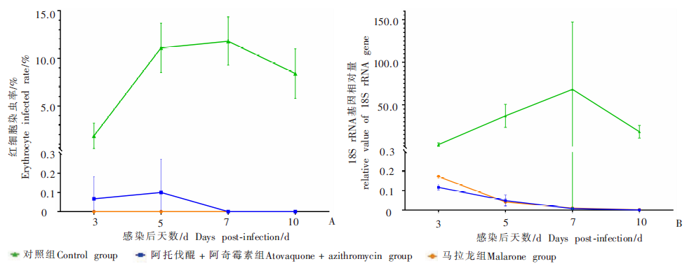
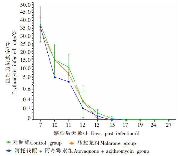
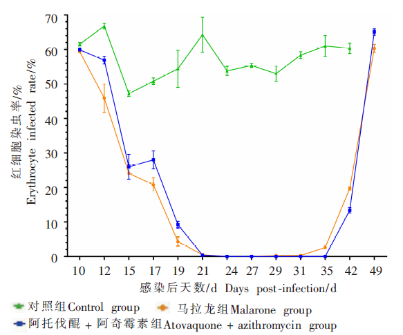
目的 以免疫状态正常的BALB/c小鼠及非肥胖糖尿病/重症联合免疫缺陷NOD/SCID小鼠为模型,对临床较常用的阿托伐醌(ATQ) + 阿奇霉素(AZM)和马拉龙进行抗田鼠巴贝虫体内药效评价。 方法 取69只BALB/c健康小鼠与15只NOD/SCID健康小鼠,每鼠接种107个感染田鼠巴贝虫的鼠红细胞。2种小鼠感染后均设置3个组,即ATQ + AZM组(195 mg/kg ATQ + 32.5 mg/kg AZM)、马拉龙组(62.5 mg/kg ATQ + 25 mg/kg氯胍,即1/4片)和对照组(5%可溶性淀粉溶液),每鼠按0.2 ml/10 g灌胃给药。BALB/c小鼠药物抑制试验中(2个用药组各12只,对照组15只),小鼠感染后4 h开始用药,连用10 d,每组于感染后1、3、5、7、10 d喂药前,随机取3只小鼠尾尖采血,采用薄血膜涂片染色镜检观察红细胞感染情况并计算红细胞染虫率(EIR),qPCR检测18S rRNA基因相对量。BALB/c小鼠药物治疗试验中(3组各10只),于感染后7 d取所有小鼠尾尖血制薄血膜染色镜检,确认感染后开始用药,连用10 d,于感染后7、10、11、12、13、15、17、19、24、27 d均采血镜检并计算EIR;于感染后27 d,每组随机取5只小鼠,采用免疫抑制剂地塞米松磷酸钠注射液(200 μl/鼠),连续腹腔注射7 d,自免疫抑制后3 d起,每天采血制薄血膜涂片染色镜检,观察复燃情况;于感染后27 d,每组随机取5只小鼠采眶窦血,将抗凝全血混匀后腹腔注射接种对应的3组健康BALB/c小鼠(每组5只),继代接种感染后7~10 d,制薄血膜涂片染色镜检并计算EIR。NOD/SCID小鼠(3组各5只)于感染后10 d开始用药,连用10 d,于感染后10、12、15、17、19、21、24、27、29、31、35、42、49 d,分别取3组小鼠尾尖血,采用薄血膜涂片染色镜检并计算EIR。应用GraphPad Prism 8对数据进行统计学分析。 结果 BALB/c小鼠药物抑制试验结果显示,ATQ + AZM及马拉龙均可有效抑制小鼠虫血症。镜检结果显示,ATQ + AZM组在感染后3 d、5 d,均仅1只小鼠查见虫体,EIR分别为(0.20 ± 0.12)%和(0.30 ± 0.17)%,感染后7 d(用药第8天),EIR降为0;马拉龙组小鼠EIR一直为0;对照组与ATQ + AZM组、马拉龙组EIR的差异均有统计学意义(F = 151.6、153.5,P < 0.05)。qPCR检测结果显示,感染后7 d,马拉龙组、ATQ + AZM组的18S rRNA基因相对量分别为0.010 2 ± 0.001 2、0.007 8 ± 0.006 6,均与对照组(68.143 8 ± 79.122 9)差异有统计学意义(F = 7.376、7.382,P < 0.05);感染后10 d(停药第1天),马拉龙组、ATQ + AZM组的18S rRNA基因相对量分别降为0.001 7 ± 0.000 9、0.000 8 ± 0.000 6,均与对照组(18.309 9 ± 7.498 6)差异有统计学意义(t = 4.229、4.229,P < 0.05)。BALB/c小鼠药物治疗试验中,对照组、ATQ + AZM组和马拉龙组的EIR均于感染后7 d达峰值,分别为(36.67 ± 10.85)%、(35.30 ± 6.46)%和(33.53 ± 7.37)%;感染后11 d(用药第5天),EIR分别降为(10.47 ± 8.02)%、(1.53 ± 0.31)%和(6.27 ± 1.01)%;感染后15 d,各组EIR逐渐趋于0。免疫抑制剂复燃试验结果显示,免疫抑制后3 d,对照组1只小鼠查见虫体;免疫抑制后5 d起,ATQ + AZM组和马拉龙组小鼠均查见虫体,出现复燃。继代接种试验结果显示,继代接种感染后7 d,ATQ + AZM组和马拉龙组均有3只受血鼠查见虫体;继代接种感染后9 d,ATQ + AZM组和马拉龙组分别有1只、2只受血鼠查见虫体;继代接种感染后10 d,ATQ + AZM组和马拉龙组受血鼠未查见虫体。ATQ + AZM组和马拉龙组NOD/SCID小鼠用药后,EIR均较快下降,从感染高峰(感染后10 d)的(59.90 ± 0.10)%和(59.37 ± 0.35)%降至感染后24 d的0,但分别于42 d、29 d又查见虫体;对照组小鼠感染后EIR在(47.20 ± 0.80)% ~(66.80 ± 0.80)%波动,于感染后45 d全部死亡,与ATQ + AZM组、马拉龙组差异有统计学意义(F = 5 505、5 984,P < 0.05)。结论 临床常用的ATQ + AZM和马拉龙对感染小鼠体内的田鼠巴贝虫增殖有一定抑制作用,但均不能完全杀灭虫体;治疗后血液仍具感染性,且在宿主免疫力低下至一定程度时虫体会复燃,呈较高红细胞染虫率。

关键词: 田鼠巴贝虫, 阿托伐醌, 阿奇霉素, 马拉龙, 药效评价, 红细胞染虫率

Abstract:

Objective To evaluate the in vivo efficacy against Babesia microti of two therapies commonly used in the clinic: a combination of atovaquone (ATQ) with azithromycin (AZM) and malarone in immune-normal BALB/c and non-obese diabetic/severe combined immunodeficient(NOD/SCID) mice models. Methods In total, 69 BALB/c and 15 NOD/SCID mice were each inoculated with 107 Babesia microti-infected erythrocytes. The mice of each immune type were divided into three groups: ATQ + AZM group (195 mg/kg ATQ + 32.5 mg/kg AZM), malarone group (62.5 mg/kg ATQ + 25 mg/kg proguanil), and control group (5% soluble starch solution), the drug was administered by gavage of 0.2 ml/10 g body weight. In the drug-suppression testing in BALB/c mice(12 mice each of two drug groups, and 15 mice in the control group), the mice received the first dose of drug 4 h after infection, and then the dosing was continued for 10 consecutive days. Three mice were randomly selected for blood sampling from tail tip before gavage administration on 1, 3, 5, 7, and 10 days post-infection. Thin blood smears were prepared for microscopy to examine B. microti infection of erythrocytes, and estimate the erythrocyte infection rate (EIR); the blood samples were tested for 18S rRNA using qPCR to examine the gene value. In the drug-therapy testing experiment with BALB/c mice (10 mice each of 3 group), tail tip blood samples were collected from all mice 7 d post-infection to determine whether the infection was established, then when the infection was confirmed, the drug administration was initiated and continued for 10 consecutive days; microscopic examination was conducted for the blood samples collected on days 10, 11, 12, 13, 15, 17, 19, 24 and 27 post-infection to estimate the EIR. On 27 d post-infection, 5 mice randomly selected from each group were intraperitoneally injected with immunosuppressant dexamethasone sodium phosphate solution (200 μl/mouse) for 7 consecutive days; starting on 3 d after immunosuppressant injection, daily blood samples were examined by staining microscopy to observe recrudescence; Orbital sinus blood samples were collected from the mice randomly selected (5 from each group), and the anticoagulated whole blood samples obtained were pooled and injected intraperitoneally to corresponding groups of normal BALB/c (5 mice/group); on 7-10 d post subinnoculation, blood samples were examined to estimate EIR. In the experiment with NOD/SCID mice (3 groups, 5 mice each group), drugs were administered from 10 d post infection and continued for 10 days. Tail tip blood samples of the mice of three groups were collected on 10, 12, 15, 17, 19, 21, 24, 27, 29, 31, 35, 42 and 49 d post-infection, and examined by staining microscopy to estimate EIR. Results In the drug-suppression test in BALB/c mice, both ATQ + ATM and malarone obviously suppressed parasitemia. Microscopic examination indicated the only one mouse in the ATQ + AZM group was found infected on 3 d and 5 d post infection, with EIR of (0.20 ± 0.12)% and (0.30 ± 0.17)% respectively, while decreased to 0 on 7 d post-infection (8 d post-dosing). EIR was 0 in all mice in the malarone group. Significant differences were observed in EIR of control vs ATQ + ATM group and control vs malarone group (F = 151.6, 153.5, P < 0.05). The relative value of 18S rRNA gene was at 0.010 2 ± 0.001 2 and 0.007 8 ± 0.006 6 in malarone and the ATQ + AZM groups, respectively on 7 d post infection, which was significantly different from the control group (68.143 8 ± 79.122 9) (F = 7.376, 7.382, P < 0.05). On 10 d post-infection (1 d upon drug withdrawal), the value of 18S rRNA gene decreased to 0.001 7 ± 0.000 9 and 0.000 8 ± 0.000 6 in the malarone and ATQ + AZM groups on day 10 post-infection, respectively, which were significantly different from the control group (t = 4.229, 4.229, P < 0.05). In the drug-therapy tests with BALB/c, EIR in control, ATQ + AZM, and malarone groups peaked on 7 d post-infection, (36.67 ± 10.85)%, (35.30 ± 6.46)%, and (33.53 ± 7.37)% respectively, and decreased to(10.47 ± 8.02)%, (1.53 ± 0.31)% and (6.27 ± 1.01)%, respectively, on 11 d post-infection (5 d upon drug withdrawal); EIR in all groups declined to 0 on 15 d post-infection. B. microti was observed in one mouse in the control group 3 d after immunosuppressant injection, and seen in both the ATQ + AZM and malarone groups 5 d after immunosuppressing, indicating that recrudescence occurred in all groups. Subinoculation experiment showed that three mice that received blood from either the ATQ + AZM or malarone groups developed parasitemia 7 d after subinoculation. One mouse and two mice showed parasitemia in the ATQ + AZM and malarone groups, respectively, 9 d after subinoculation, and no parasite was observed in any mice from 10 d post subinoculation. In the test with NOD/SCID mice, the EIR of ATQ + AZM and malarone groups rapidly reduced after treatment from its peak value on 10 d post infection [(59.90 ± 0.10) % and (59.37 ± 0.35)%, respectively] to 0 on 24 d post-infection, whereas B. microti was observed again on day 29 and 42 post infection. EIR in the control group fluctuated between (47.20 ± 0.80)% and (66.80 ± 0.80)%, and all mice died on 45 d post-infection, of which the process was significantly different from ATQ + ATM, and malarone group (F = 5 505 and 5 984, respectively, P < 0.05). Conclusion Both ATQ + ATM and malarone that commonly used in clinic showed suppressive effect to some extent on the proliferation of B. microti in infected mice, but could not clear up all the parasites; the blood of mice remains infective after drug treatment and recrudescence may occur in immunocompromised hosts, causing high infection rate of erythrocyte.

Key words: Babesia microti, Atovaquone, Azithromycin, Malarone, Drug efficacy evaluation, Erythrocyte infected rate

中图分类号: