中国寄生虫学与寄生虫病杂志 ›› 2020, Vol. 38 ›› Issue (6): 710-717.doi: 10.12140/j.issn.1000-7423.2020.06.006

• 论著 • 上一篇    下一篇

传播阻断疫苗候选抗原Pb280在伯氏疟原虫中的表达及功能研究

吴宇迪, 刘飞, 杨帆, 曹雅明*   

  1. 中国医科大学基础医学院免疫学教研室,沈阳 110122
  • 收稿日期:2020-03-04 出版日期:2020-12-30 发布日期:2021-01-12
  • 通讯作者: 曹雅明
  • 基金资助:
    国家自然科学基金(81761128017)

Expression and function of a potential antigen Pb280 as transmission-blocking vaccine in Plasmodium berghei

WU Yu-di, LIU Fei, YANG Fan, CAO Ya-ming*   

  1. Department of Immunology, College of Basic Medical Sciences, China Medical University, Shenyang 110122, China
  • Received:2020-03-04 Online:2020-12-30 Published:2021-01-12
  • Contact: CAO Ya-ming
  • Supported by:
    National Nature Science Foundation of China(81761128017)

摘要:

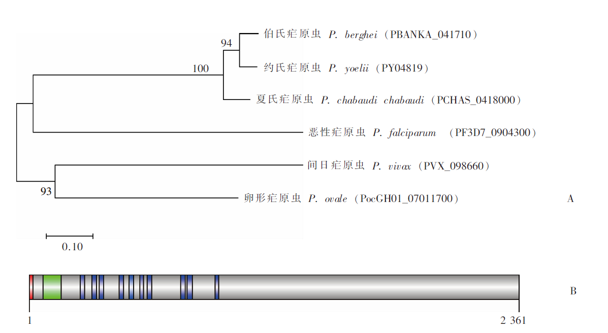
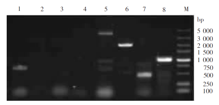
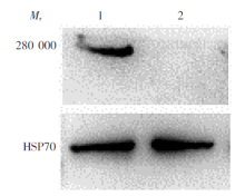
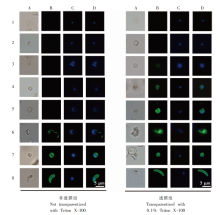
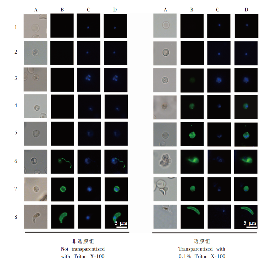
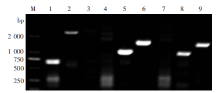
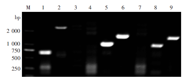
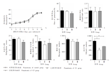
目的 探讨传播阻断疫苗候选抗原Pb280(PBANKA_041710)在伯氏疟原虫中的表达情况及基因功能。 方法 从NCBI数据库获取不同疟原虫种属Pb280氨基酸同源序列,用MEGA7构建系统进化树。用SMART在线网站预测Pb280蛋白结构域。昆明小鼠经尾静脉注射1 × 10⁶Pb感染红细胞(iRBC),当小鼠原虫血症达3%~5%时取眼球血进行体外培养并分离裂殖体。将带血凝素(HA)的Pb280标签质粒(Pb280HA)和Pb280敲除质粒(Pb280KO)线性化后分别转染裂殖体,用转染后裂殖体注射感染小鼠(1 × 10⁷个裂殖体/鼠),PCR鉴定感染小鼠中的转基因疟原虫,对转基因疟原虫进行单克隆筛选,获得1株Pb280HA标签虫株和2株Pb280KO虫株(分别命名为Pb280HA型、Pb280KO-C1型、Pb280KO-C2型虫株)。蛋白质免疫印迹分析(Western blotting)和间接免疫荧光试验(IFA)分析Pb280在伯氏疟原虫中的表达情况。取9只雌性昆明小鼠随机分为基因敲除C1组(C1组)、基因敲除C2组(C2组)和对照组,每组3只,每鼠分别经尾静脉注射5 × 10⁶Pb280KO-C1型、Pb280KO-C2型和野生型(WT)伯氏疟原虫。感染后第3~10天,每天取小鼠尾静脉血进行血涂片检测,计算原虫血症;计算感染后第3天雌雄配子体比及配子体率。感染后第3天取小鼠尾静脉血与动合子培养基混合培养15 min后计数雄配子体出丝中心数、雌雄配子结合数、雌配子数,继续培养24 h后计数动合子数。原虫血症、配子体率和雌雄配子体比的比较采用χ2检验,其余各组数据之间的比较采用单因素方差分析。 结果 系统进化树显示,Pb280与约氏疟原虫PY04819的亲缘关系最近。SMART预测结果显示,Pb280含有一个N端信号肽、10个跨膜区和1个生长因子受体结构域。Western blotting分析显示,Pb280蛋白在伯氏疟原虫中有表达,相对分子质量(Mr)约280 000。IFA检测结果显示,在配子体到动合子的发育过程中,Pb280从胞浆向质膜表面分泌。基因功能分析结果显示,感染后第3天,3组小鼠原虫血症均约为15%,感染后第10天均达60%以上,C1组、C2组与对照组的差异无统计学意义(P > 0.05)。感染后第3天,C1组配子体率、雌雄配子体比、平均每10个视野配子体出丝中心数量、雌雄配子结合数和雌配子数分别为39.50‰、1.65、(21.63 ± 4.03)个、(12.50 ± 8.02)个和(930.00 ± 79.20)个,C2组分别为34.50‰、1.71、(18.25 ± 5.85)个、(13.75 ± 9.54)个和(885.00 ± 130.11)个,对照组分别为41.50‰、1.74、(21.44 ± 4.73)个、(15.31 ± 8.06)个和(1 018.50 ± 58.69)个,3组比较差异均无统计学意义(P > 0.05)。C1组动合子形成数量为(410.00 ± 67.88)个,C2组为(557.50 ± 2.12)个,对照组为(782.00 ± 41.01)个,C1和C2组动合子数量均少于对照组(P < 0.05),C1组和C2组差异无统计学意义(P > 0.05)。 结论 Pb280在疟原虫种属中保守,在伯氏疟原虫的裂殖体、配子体及动合子阶段均有表达,敲除该基因可导致动合子数量减少。

关键词: 疟疾, 伯氏疟原虫, 传播阻断疫苗, Pb280

Abstract:

Objective To investigate the expression and function of a transmission-blocking vaccine candidate Pb280 (PBANKA_041710) in Plasmodium berghei. Methods Homologous sequences of Plasmodium species were downloaded from NCBI and compared using MEGA7. The domains of Pb280 were predicted by SMART. Ten female Kunming (KM) mice aged 6-8 weeks were randomly selected, injected with 1 × 10⁶ P. berghei-infected red blood cells (iRBC) through tail vein. When the parasitemia in the mice reached 3%-5%, orbital blood was obtained and cultured in vitro for isolation of schizonts. The hemagglutinin (HA) tagged plasmid (Pb280HA) and Pb280 knockout plasmid (Pb280KO) were linearized and transfected to mature schizonts, which were then injected in the mice (1 × 10⁷ schizonts/mouse) through tail vein; transgenic parasites in the infected mice was identified by PCR, and underwent monoclonal screening. As a result, one strain of Pb280HA and two strains of Pb280KO (Pb280KO-C1 and Pb280KO-C2) were obtained. The expression of Pb280 in P. berghei was analyzed by Western blotting and indirect immunofluorescence assay (IFA). Another nine female KM mice were randomly divided into 3 groups (n = 3): Pb280KO-C1 group (C1 group), Pb280KO-C2 group (C2 group) and control group. Each mouse was injected with 5 × 10⁶ Pb280KO-C1, Pb280KO-C2 or wild-type (WT) P. berghei through tail vein. From day 3 to day 10 post-infection, mice tail vein blood was sampled for monitoring daily parasitemia by examining blood smears. On day 3 after infection, gametocytemia and female/male gametes ratio were calculated; mice tail vein blood was mixed with ookinete culture medium for 15 min to count the number of exflagellation centers, number of male-female integrating and number of macrogametes followed by further culture for 24 h to count the number of ookinetes. The comparisons of parasitemia, gametocytemia and female/male gamete ratio were performed by Chi-square test, and other data were analyzed with one-way ANOVA. Results Phylogenetic tree constructed by MEGA7 showed that Pb280 was genetically most close to the PY04819 of P. yoelii. SMART prediction showed that Pb280 contained an N-terminal signal peptide, 10 transmembrane domains and 1 growth factor receptor domain. Western blotting demonstrated that Pb280 protein expression occurred in P. berghei, with a relative molecular weight of 280 000. IFA results showed that Pb280 was secreted from cytoplasm to plasma membrane during the development from gametocyte to ookinete. Gene function analysis revealed that on day 3 after infection, the three groups had parasitemia of about 15%, which reached over 60% on day 10 post-infection. The parasitemia rate in the group C1 and the C2 did not differ significantly from that in the control group (P > 0.05). On day 3 post-infection, the gametocytemia rate, gamete sex ratio, number of exflagellation centers per 10 fields of view, number of male-female integrating and number of macrogametes in group C1 were 39.50‰, 1.65, 21.63 ± 4.03, 12.50 ± 8.02, and 930.00 ± 79.20, respetively; those in the group C2 were 34.50‰, 1.71, 18.25 ± 5.85, 13.75 ± 9.54, and 885.00 ± 130.11, respetively; and those in the control group were 41.50‰, 1.74, 21.44 ± 4.73, 15.31 ± 8.06, and 1 018.50 ± 58.69. There were no statistically significant differences among the three groups (P > 0.05). The numbers of ookinetes in the C1, C2 and control groups were 410.00 ± 67.88, 557.50 ± 2.12 and 782.00 ± 41.01, respectively. The numbers of ookinetes in the C1 and C2 groups did not differ significantly, but both were lower than that in the control group (P < 0.05). Conclusion Pb280 is genetically conserved in Plasmodium spp. and expressed in schizont, gametocyte and ookinete stages of P. berghei. Knockout Pb280 may result in the decrease of ookinete formation.

Key words: Malaria, Plasmodium berghei, Transmission-blocking vaccines, Pb280

中图分类号: