中国寄生虫学与寄生虫病杂志 ›› 2020, Vol. 38 ›› Issue (5): 611-618.doi: 10.12140/j.issn.1000-7423.2020.05.013

• 论著 • 上一篇    下一篇

多房棘球蚴感染小鼠脾CD4+ T细胞亚群及其功能耗竭的变化

侯昕伶1,2(), 李玲慧3, 李亮1, 李静2, 王慧1,2, 邵英梅4, 张传山1,2,*()   

  1. 1 新疆医科大学第一附属医院临床医学研究院,省部共建中亚高发病成因与防治国家重点实验室,新疆包虫病基础医学重点实验室,乌鲁木齐 830054
    2 新疆医科大学基础医学院,乌鲁木齐 830054
    3 新疆农业大学动物医学学院,乌鲁木齐 830054
    4 新疆医科大学第一附属医院,消化血管外科中心肝胆包虫科,乌鲁木齐 830054
  • 收稿日期:2020-05-09 出版日期:2020-10-30 发布日期:2020-11-12
  • 通讯作者: 张传山
  • 作者简介:侯昕伶(1995-)女,硕士研究生,从事寄生虫感染与免疫。E-mail:465778824@qq.com
  • 基金资助:
    省部共建中亚高发病成因与防治国家重点实验室开放课题(SKL-HIDCA-2019-2);国家自然科学基金(81760368);国家自然科学基金(81560330);新疆维吾尔自治区高校科研计划项目(XJEDU2020Y024)

Changes in subsets and functional exhaustion of CD4+ T cells in spleens of mice infected with Echinococcus multilocularis

HOU Xin-ling1,2(), LI Ling-hui3, LI Liang1, LI Jing2, WANG Hui1,2, SHAO Ying-mei4, ZHANG Chuan-shan1,2,*()   

  1. 1 Clinical Medicine Institute, State Key Laboratory of Pathogenesis, Prevention and Treatment of High Incidence Diseases in Central Asia, Xinjiang Key Laboratory of Echinococcosis, the First Affiliated Hospital of Xinjiang Medical University, Urumqi 852204, China
    2 Basic Medical College, Xinjiang Medical University, Urumqi 852204, China
    3 College of Animal Medicine, Xinjiang Agricultural University, Urumqi 852204, China
    4 Department of Hepatic Hydatid and Hepatobiliary Surgery, Digestive and Vascular Surgery Centre, the First Affiliated Hospital of Xinjiang Medical University, Urumqi 852204, China
  • Received:2020-05-09 Online:2020-10-30 Published:2020-11-12
  • Contact: ZHANG Chuan-shan
  • Supported by:
    Open Project of State Key Laboratory of Pathogenesis, Prevention and Treatment of High Incidence Diseases in Central Asia(SKL-HIDCA-2019-2);National Natural Science Foundation of China(81760368);National Natural Science Foundation of China(81560330);Xinjiang Uygur Autonomous Region University Scientific Research Project(XJEDU2020Y024)

摘要:

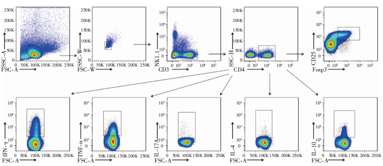
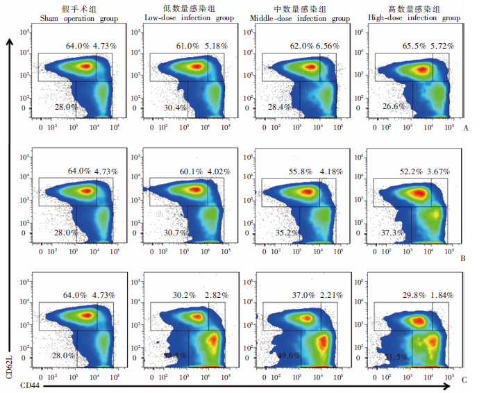
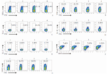
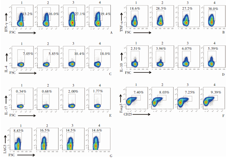
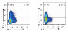
目的 研究不同数量多房棘球蚴感染对小鼠脾CD4+ T细胞亚群及其免疫功能的影响。方法 60只C57BL/6小鼠随机分为4组,每组15只,分别为假手术组、低数量感染组(50个原头节)、中数量感染组(500个原头节)和高数量感染组(2 000个原头节)。小鼠麻醉后经肝门静脉部位穿刺,注射不同数量原头节,假手术组注射等量生理盐水。于感染后2、12和24周各组分别取5只小鼠,取脾组织研磨分离淋巴细胞。流式细胞术检测各组小鼠脾CD4+ T细胞记忆表型、不同亚群比例、免疫抑制性分子淋巴细胞活化蛋白3(LAG3)表达。采用GraphPad Prism 6.0软件进行作图和统计学分析。结果 感染后2周,低数量和中数量感染组小鼠脾CD4+IFN-γ+ T细胞比例分别为(7.54 ± 1.44)%、(7.58 ± 3.17)%,高于假手术组的(3.52 ± 1.03)%(P < 0.05);CD4+TNF-α+ T细胞比例分别为(39.34 ± 4.19)%、(39.53 ± 10.74)%,高于假手术组(22.62 ± 1.50)% (P < 0.01)。感染后12周,低数量和中数量感染组小鼠脾CD4+IFN-γ+ T细胞比例分别为(16.52 ± 0.77)%、(22.98 ± 4.32)%,高于假手术组(16.88 ± 2.49)%(P < 0.05);CD4+TNF-α+ T细胞比例分别为(27.26 ± 2.12)%、(28.36 ± 5.24)%,高于假手术组(19.72 ± 3.87)% (P < 0.05);CD4+IL17A+ T细胞比例分别为(10.70 ± 1.81)%、(11.52 ± 2.68)%,高于假手术组(5.40 ± 1.32)% (P < 0.01);同时,低数量和中数量感染组小鼠脾CD4+IL-4+ T细胞比例分别为(2.87 ± 0.84)%、(3.50 ± 0.77)%,高于假手术组(1.75 ± 0.83)% (P < 0.01);CD4+IL-10+ T细胞比例分别为(4.63 ± 0.78)、(7.09 ± 2.42)%,高于假手术组(3.03 ± 0.79)% (P < 0.01)。感染后24周,中数量、高数量感染组小鼠脾CD4+IFN-γ+ T、CD4+TNF-α+ T、CD4+IL-4+ T、CD4+IL-10+ T和CD4+IL17A+ T细胞的比例均高于假手术组(P < 0.05),且高数量组小鼠脾Treg细胞的比例高于假手术组(P < 0.01),各感染组小鼠脾效应记忆性CD4+ T细胞比例高于假手术组;各感染组小鼠脾CD4+LAG3+ T细胞比例分别为(16.45 ± 4.89)%、(14.54 ± 4.96)%、(14.62 ± 2.43)%,高于假手术组(8.43 ± 3.46)%(P < 0.05)。感染后24周, 高数量组小鼠脾CD4+ T细胞中分泌IFN-γ和TNF-α的LAG3阳性群细胞比例分别为(1.67 ± 0.66)%、(0.69 ± 0.27)%,低于阴性群的(5.11 ± 1.81)%、(31.7 ± 12.1)%(P < 0.01)。结论 低、中数量多房棘球蚴感染后,小鼠可能利用T1型和T17型免疫应答优势对虫体起到杀伤和清除;而高数量感染诱导脾T1/T2型和T17/Treg型免疫应答失衡,以及CD4+ T细胞上调LAG3分子表达,导致功能耗竭,造成棘球蚴慢性寄生。

关键词: 多房棘球蚴病, 脾, CD4+ T细胞, 功能耗竭, 淋巴细胞活化蛋白3

Abstract:

Objective To investigate the effects of different doses of Echinococcus multilocularis infection on splenic CD4+ T cell subsets and immune functions in mice. Methods Sixty female C57BL/6 mice were randomly divided into 4 groups, 15 in each group, including sham operation group (saline), low-dose infection group (50 protoscoleces/mouse), medium-dose infection group (500 protoscoleces/mouse), and high-dose infection group (2 000 protoscoleces/mouse). Mice were inoculated via the hepatic portal vein under anaesthetia with different doses of protoscoleces in saline, whereas the control mice were injected with the same volume of saline. Spleens were taken from 5 mice of each group at 2, 12, and 24 weeks after infection, and ground to isolate lymphocytes. Flow cytometry was performed to detect the memory phenotype, the proportions of different subsets, and LAG3 expression in splenic CD4+ T cells in different experimental groups. Statistical analysis was performed using Graphad Prism 6.0 software. Results At 2 weeks after infection, the proportions of splenic CD4+IFN-γ+ T cells in the low- and medium-dose groups were (7.54 ± 1.44)% and (7.58 ± 3.17)%, respectively, which were higher than that in the sham operation group [(3.52 ± 1.03)%, P < 0.05]; and the proportions of splenic CD4+TNF-α+ T cells were (39.34 ± 4.19)% and (39.53 ± 10.7) %, respectively, which were higher than that of the sham operation group [(22.62 ± 1.50)%, P < 0.01]. At 12 weeks after infection, the proportions of splenic CD4+IFN-γ+ T cells in the low- and medium-dose groups were (16.52 ± 0.77)% and (22.98± 4.32)%, respectively, which were higher than that in the sham operation group [(16.88 ± 2.49)%, P < 0.05]; the proportions of splenic CD4+TNF-α+ T cells were (27.26 ± 2.12)% and (28.36 ± 5.24)%, respectively, which were higher than that of the sham operation group [(19.72 ± 3.87)%, P < 0.05]; and the proportions of splenic CD4+IL-17A+ T cells were (10.70 ± 1.81)% and (11.52 ± 2.68)%, respectively, which were higher than that of the sham operation group [(5.40 ± 1.32)%, P < 0.01]. In addition, the proportions of splenic CD4+IL-4+ T cells in the low- and medium-dose groups were (2.87 ± 0.84)% and (3.50 ± 0.77)%, respectively, which were higher than that of the sham operation group (1.75 ± 0.83)% (P < 0.01); and the proportions of splenic CD4+IL-10+ T cells were (4.63 ± 0.78)% and(7.09 ± 2.42)%, respectively, which were higher than that of the sham operation group [(3.03 ± 0.79)%, P < 0.01]. At 24 weeks after infection, the proportions of splenic CD4+IFN-γ+ T cells, CD4+TNF-α+ T cells, CD4+IL-4+ T cells, CD4+IL-10+ T cells and CD4+IL-17A+ T cells in the medium- and high-dose groups were all higher than those in the sham operation group (P < 0.05), and the proportion of splenic Treg cells in the high-dose group was higher than that in the sham operation group (P < 0.01). Moreover, at 24 weeks, the proportions of splenic CD4+ Treg cells in the three infection groups were all higher than that in the sham operation group; and the proportions of splenic CD4+LAG3+ T cells in the three infection groups were (16.45 ± 4.89)%, (14.54 ± 4.96)%, and (14.62 ± 2.43)%, respectively, which were higher than that of the sham operation group (8.43 ± 3.46)% (P < 0.05). At 24 weeks after infection, the proportions of LAG3-positive CD4+ T cells sereting IFN-γ and TNF-α in the high dose group were (1.67 ± 0.66)% and (0.69 ± 0.27)%, which were significantly lower than those LAG3-negative CD4+ T cells[(5.11 ± 1.81)% and(31.7 ± 12.1)%, P < 0.01]. Conclusion After infected with low and medium doses of E. multilocularis, the mice may make advantages of T1 and T17 immune responses to kill and clear the worms, while infection with a high dose of E. multilocularis may lead to imbalance of T1/T2 and T17/Treg type immune responses, and the occurance of CD4+ T cells up-regulating the expression of LAG3, resulting in the exhaustion of CD4+ T cells and causing chronic alveolar echinococcosis.

Key words: Alveolar echinococcoisis, Spleen, CD4+ T cells, Functional exhaustion, Lymphocyte-activation gene 3

中图分类号: