中国寄生虫学与寄生虫病杂志 ›› 2019, Vol. 37 ›› Issue (6): 676-680.doi: 10.12140/j.issn.1000-7423.2019.06.010

• 论著 • 上一篇    下一篇

肝毛细线虫体外培养及小鼠感染模型的建立

张雅兰1(), 朱岩昆1, 高丽君1, 王磊2, 邓艳1, 陈伟奇1, 许汴利1, 蔺西萌1, 张红卫1,*()   

  1. 1河南省疾病预防控制中心,郑州 450016
    2 首都医科大学附属北京友谊医院,北京热带医学研究所,热带病防治北京市重点实验,北京 100050
  • 收稿日期:2019-04-09 出版日期:2019-12-30 发布日期:2019-12-31
  • 通讯作者: 张红卫
  • 作者简介:

    作者简介:张雅兰(1988-),女,硕士,主管技师,主要从事寄生虫病预防控制工作。E-mail:zylxiaokedou@163.com

  • 基金资助:
    国家自然科学基金(No. 81702018);河南省医学科技攻关计划项目(No. 201702280)

Establishment of a mouse model of Capillaria hepatica infection

Ya-lan ZHANG1(), Yan-kun ZHU1, Li-jun GAO1, Lei WANG2, Yan DENG1, Wei-qi CHEN1, Bian-li XU1, Xi-meng LIN1, Hong-wei ZHANG1,*()   

  1. 1 Henan Center for Disease Control and Prevention, Zhengzhou 450016, China
    2 Beijing Institute of Tropical Medicine, Beijing Key Laboratory for Prevention and Treatment of Tropical Diseases, Beijing Friendship Hospital, Capital Medical University, Beijing 100050, China
  • Received:2019-04-09 Online:2019-12-30 Published:2019-12-31
  • Contact: Hong-wei ZHANG
  • Supported by:
    Supported by National Natural Science Foundation of China (No. 81702018) and Medical Science and Technology Project of Henan Province of China (No. 201702280)

摘要:

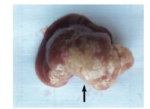
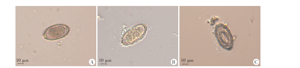
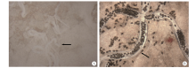
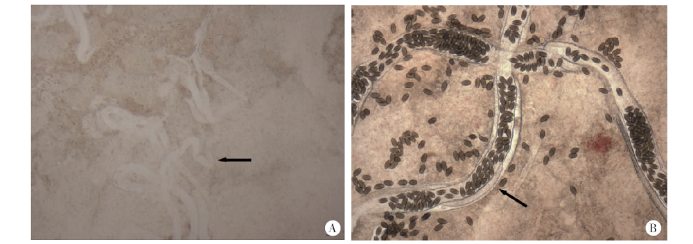
目的 建立肝毛细线虫感染的小鼠模型,为肝毛细线虫病的研究奠定基础。 方法 在洛阳鼠密度较高地区捕鼠,以人工消化法收集肝毛细线虫阳性鼠肝内的虫卵。将虫卵以不同密度(1 000、5 000和10 000个/孔)分为3组,每组2孔,于28 ℃培养至感染期,观察并计数不同培养时长时(18、21、23、25和29 d)感染期虫卵的比例。将感染期虫卵分为两组,分别置于蒸馏水和人工胃液中37 ℃培养,定期观察有无幼虫孵出。将4周龄雄性昆明小鼠分为8组,每组3只,按5、10、20、60、100、200、500和800个虫卵/只将感染期肝毛细线虫卵通过灌胃感染小鼠,观察小鼠感染后存活情况及是否能成功感染肝毛细线虫。另以16只昆明小鼠作为发育观察组,以60个虫卵/只的感染,分别于感染后14、18、21、29、35、55、90和365 d取2只小鼠处死,解剖取肝脏镜检,观察肝毛细线虫在小鼠体内的发育及繁殖情况。 结果 共捕鼠17只,肝毛细线虫感染阳性鼠5只,阳性率为5/17;所有阳性鼠均为褐家鼠,褐家鼠阳性率为5/8。新鲜收集的肝毛细线虫虫卵内的卵细胞多处于Ⅲ或Ⅳ细胞期,培养1 d后分裂加速,至第16天开始出现感染期虫卵。1 000、5 000和10 000个/孔体外培养18、23、25和29 d时虫卵中感染期虫卵的比例分别为:39.6%(67/169)、35.2%(45/128)、21.4%(98/458),74.0%(148/200)、75.1%(411/547)、60.9%(340/558),88.0%(125/142)、89.2%(140/157)、79.3%(168/212)和94.9%(131/138)、97.0%(254/262)、88.6%(140/158),各时间点的组间差异均有统计学意义(P < 0.05),且10 000个/孔密度条件下的比例均最低。分别在蒸馏水和人工胃液中培养感染期虫卵均无幼虫孵出。感染剂量在200个/只及以下的小鼠均可成功感染肝毛细线虫且未出现死亡;500和800个/只的剂量组小鼠均出现死亡。发育观察组于感染后14 d鼠肝压片查见肝毛细线虫幼虫,未见雌雄分化;感染后18、21和29 d,肝压片可见肝毛细线虫孕虫;感染后35和55 d均见大量肝毛细线虫虫卵及虫体片段;感染后90 、365 d仅见肝毛细线虫虫卵,无虫体残余组织。 结论 体外培养可获得感染期肝毛细线虫虫卵,小鼠可作为肝毛细线虫感染的动物模型。

关键词: 肝毛细线虫, 动物模型, 小鼠

Abstract:

Objective To establish a mouse infection model of Capillaria hepatica to facilitate the research on hepatic capillariasis. Methods The C. hepatica infected rats were collected from endemic area in Luoyang, Henan. The C. hepatica eggs were collected from the liver of an infected rat by 1% pepsin digestion. The collected eggs were divided into 3 groups with 1 000, 5 000 and 10 000 per well in duplicate, then cultured in vitro at 28 ℃ in an incubator. The eggs were observed under microscope for their embryonated rate at Day 18, 21, 23, 25, 29. The fully embryonated eggs were divided into two groups and incubated in distilled water and artificial gastric juice respectively under 37 ℃. The hatched larvae were observed. Three male Kunming mice with 4 weeks old in each group were inoculated orally with 5, 10, 20, 60, 100, 200, 500 and 800 C. hepatica embryonated eggs. The survival rate of each group was observed as evidence of successful infection. Another sixteen mice were infected orally with 60 C. hepatica embryonated eggs to systemically observe the infection and development of C. hepatica worms and eggs in livers at Day 14, 18, 21, 29, 35, 55, 90, 365 post infection by necropsying 2 mice at each time point. Results Seventeen rats were captured from endemic area Luoyang, Henan, and five identified to be infected with C. hepatica with infection ratio of 5/17. Rattus norvegicus rat was the only species infected with C. hepatica with infection rate of 5/8. Most of C. hepatica eggs observed in the livers of infected rats were at Ⅲ to Ⅳ cell divided stage. After being cultured in vitro, the cell in collected C. hepatica eggs started to develop and divide to multiple cells. The embryonated eggs could be seen on Day 16 of culture. The rates of embryonated eggs in the culture groups of 1 000, 5 000 and 10 000 eggs per well were 39.6% (67/169), 35.2% (45/128) and 21.4% (98/458) on Day 18; 74.0% (148/200), 75.1% (411/547) and 60.9% (340/558) on Day 23; 88.0% (125/142), 89.2% (140/157) and 79.3% (168/212) on Day 25; and 94.9% (131/138), 97.0% (254/262) and 88.6% (140/158) on Day 29; respectively, with statistical significance between each culture time points (P < 0.05). No larva was hatched from embryonated eggs either in distilled water or in artificial gastric juice. The Kunming mice were successfully infected with 200 or less embryonated eggs of C. hepatica and all survived during the infection. The mice could not survive with infection of more than 500 eggs. In the groups of mice infected with 60 C. hepatica embryonated eggs, the larvae of C. hepatica could be observed in the liver tissue on Day 14. The matured and pregnant female adult worms were observed on Day 18. The adult worms started to die and the unembryonated eggs were laid into liver tissue on Day 35. All worms degenerated and only eggs were observed in liver tissue on Day 90 and 365 post infection. Conclusion The embryonated eggs of C. hepatica can be acquired by in vitro culture of eggs collected from infected rat and C. hepatic infection model was successfully established in Kunming mice with less than 200 embryonated infective eggs.

Key words: Capillaria hepatica, Animal model, Mice

中图分类号: