中国寄生虫学与寄生虫病杂志 ›› 2019, Vol. 37 ›› Issue (5): 571-576.doi: 10.12140/j.issn.1000-7423.2019.05.011

• 论著 • 上一篇    下一篇

阿霉素体外抗细粒棘球蚴原头节作用研究

田春艳1(), 陈蓓2,3, 卢帅2,3, 文丽梅2,3, 赵军2,3, 郑璇1, 库尔班泥沙·阿马洪1, 高旖1, 王建华2,3,*()   

  1. 1 新疆医科大学 1药学院
    2 第一附属医院药学部
    3 第一附属医院,省部共建中亚高发病成因与防治国家重点实验室,乌鲁木齐 830054
  • 收稿日期:2019-03-25 出版日期:2019-10-30 发布日期:2019-11-07
  • 通讯作者: 王建华
  • 作者简介:

    作者简介:田春艳(1993-),女,硕士,从事棘球蚴病新药及作用机制研究。E-mail:tcy1416@163.com

  • 基金资助:
    国家自然科学基金(No. 81860666,No. 81560607);省部共建中亚高发病成因与防治国家重点实验室开放课资助项目(No. SKL-HIDCA-2017-11,No. SKL-HIDCA-2017-Y7);新疆维吾尔自治区药学会项目(No. YXH201704)

Toxicity of doxorubicin on Echinococcus granulosus protoscoleces in vitro

Chun-yan TIAN1(), Bei CHEN2,3, Shuai LU2,3, Li-mei WEN2,3, Jun ZHAO2,3, Xuan ZHENG1, Kuerbannisha·AMAHONG1, Yi GAO1, Jian-hua WANG2,3,*()   

  1. 1 College of Pharmaceutical Sciences, Xinjiang Medical University, Urumqi 830054, China
    2 Department of Pharmacy, The First Affiliated Hospital of Xinjiang Medical University,Urumqi 830054, China
    3 The State Key Laboratory of Pathogenesis, Prevention and Treatment of Diseases Prevalent in Central Asia, The First Affiliated Hospital of Xinjiang Medical University, Urumqi 830054, China
  • Received:2019-03-25 Online:2019-10-30 Published:2019-11-07
  • Contact: Jian-hua WANG
  • Supported by:
    Supported by the National Natural Science Foundation of China (No. 81860666, No. 81560607), the Ministry-Provincial Cooperation for the Establishment of the State Key Laboratory on the Causes and Control of High Incidence of Disease in Central Asia (No. SKL-HIDCA-2017-11, No. SKL-HIDCA-2017-Y7) and the Project of Xinjiang Uygur Autonomous Region Medical Association (No. YXH201704)

摘要:

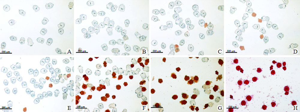
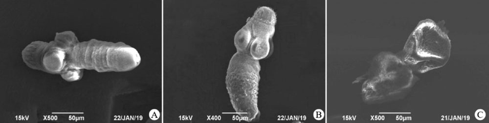
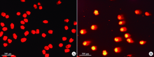
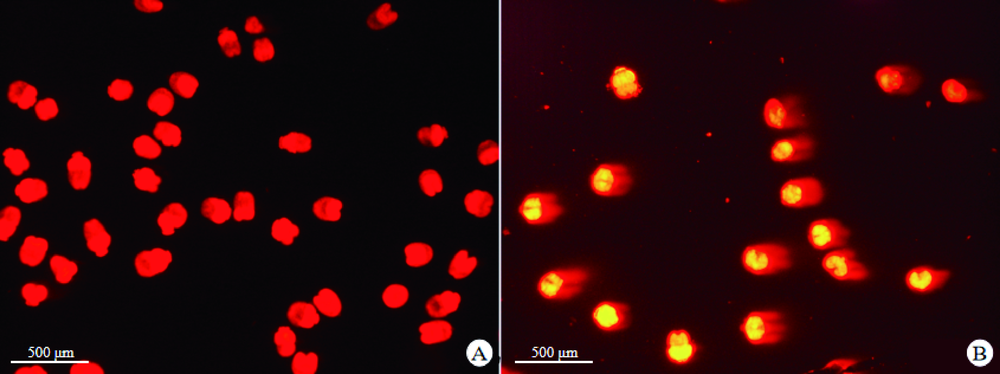
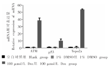
目的 研究阿霉素(Dox)对细粒棘球蚴原头节的体外抑制作用。方法 4 800个原头节随机分为空白对照组、1% DMSO组、Dox组(25、50、100、200、400、600 μmol/L),每组200个原头节,设3个平行组,体外干预24 h。伊红拒染实验检测Dox干预后原头节活力,统计存活率。扫描电镜观察Dox干预后原头节表面超微结构变化。单细胞凝胶电泳实验检测Dox干预后原头节DNA损伤程度。实时荧光定量PCR(qRT-PCR)检测Dox干预后DNA损伤相关基因共济失调毛细血管扩张症突变激酶(ATM)、p53、DNA拓扑异构酶2A(Topo2a)的mRNA表达水平。结果 伊红拒染实验结果显示,空白对照组、1% DMSO组、25、50、100、200、400、600 μmol/L Dox组细粒棘球蚴原头节的存活率分别为(99.0 ± 0.5)%、(98.6 ± 0.3)%、(96.0 ± 1.4)%、(80.3 ± 4.8)%、(75.6 ± 6.2)%、(53.2 ± 3.0)%、(26.4 ± 8.1)%和0,IC50为(267.9 ± 7.1)μmol/L。与空白对照组相比,1% DMSO组、25 μmol/L Dox组原头节存活率差异无统计学意义(P > 0.05),50、100、200、400、600 μmol/L Dox组原头节存活率差异有统计学意义(P < 0.01)。扫描电镜结果显示,空白对照组、1% DMSO组原头节表面光滑,头节呈球形或椭圆形,顶突完整,微毛清晰可见;400 μmol/L Dox组原头节吸盘变形,体部塌陷严重,顶突轻微变形,微毛明显脱落,虫体表层发生皱缩。单细胞凝胶电泳实验结果显示,100 μmol/L Dox组原头节出现拖尾现象,彗星尾距(OTM)为16.6 ± 1.7,与空白对照组(0.1 ± 0.0)相比,差异有统计学意义(P < 0.01)。qRT-PCR结果显示,1% DMSO组原头节ATM、p53和Topo2a mRNA相对表达量分别为1.0 ± 0.1、1.1 ± 0.1、1.3 ± 0.9,与空白对照组(1.0 ± 0.1、1.0 ± 0.4、1.0 ± 0.1)相比,差异无统计学意义(P > 0.05)。100 μmol/L Dox组原头节ATM、p53和Topo2a mRNA相对表达量分别为38.6 ± 3.5、10.0 ± 2.5、54.0 ± 0.8,与空白对照组相比,差异有统计学意义(P < 0.05)。结论 Dox具有体外抗细粒棘球蚴原头节作用,可引起虫体DNA损伤,可能与ATM、p53、Topo2a的mRNA相对表达量变化相关。

关键词: 阿霉素, 细粒棘球蚴原头节, DNA损伤, 共济失调毛细血管扩张症突变激酶, p53, Topo2a

Abstract:

Objective To evaluate the toxicity of doxorubicin (Dox) on Echinococcus granulosus protoscoleces in vitro. Methods Total 200 E. granulosus protoscoleces in each group were incubated with different concentration of doxorubicin (25, 50, 100, 200, 400, 600 μmol/L) in triplicate for 24 hours in vitro. Other two groups were incubated with medium(blank) or 1% DMSO controls. The viability of protoscoleces was determined by eosin staining and survival rate was calculated. The structural damage of treated protoscoleces was observed under scanning electron microscope (SEM). Single cell gel electrophoresis was used to detect DNA damage in treated protoscoleces. Real-time quantitative PCR (qRT-PCR) was used to detect the mRNA expression levels of ataxia telangiectasia mutated gene (ATM), p53 and DNA topoisomerase 2-alpha (Topo2a) in treated protoscoleces. Results Based on the eosin dye staining, the survival rates of treated protoscoleces were (96.0 ± 1.4)%, (80.3 ± 4.8)%, (75.6 ± 6.2)%, (53.2 ± 3.0)%, (26.4 ± 8.1)% and 0 when the concentrations of Dox were 25, 50, 100, 200, 400, 600 μmol/L, respectively, compared to that of protoscoleces incubated with blank control [(99.0 ± 0.5)%] and 1% DMSO (98.6 ± 0.3)%], with significant difference when incubated concentration is over 25 μmol/L (P < 0.01). The Dox IC50 on protoscoleces was (267.92 ± 7.14) μmol/L. SEM results showed that E. granulosus protoscoleces in the blank control group and 1% DMSO group had smooth surface with a spherical or elliptic shaped scolex and complete apical structure. There were clearly visible microtricles on the surface. When incubated with 400 μmol/L Dox, the protoscoleces scolex was distorted and damaged and whole body collapsed. The apical structure slightly distorted. The microtriches on the surface were lost obviously. The body surface was shrinked. The single cell gel electrophoresis showed trailed cell when the concentration of Dox reached to 100 μmol/L, indicating the cell damage. The olive tail moment (OTM) was 16.6 ± 1.7 for cells treated with 100 μmol/L of Dox, which was significantly longer compared with the blank control group(0.1 ± 0.0) (P < 0.01). The qRT-PCR results showed that the relative expressions of ATM, p53 and Topo2a mRNA in 1% DMSO group were 1.0 ± 0.1, 1.1 ± 0.1 and 1.3 ± 0.9, respectively, with no statistical significance compared with the blank control group (1.0 ± 0.1, 1.0 ± 0.4, 1.0 ± 0.1, respectively) (P > 0.05). However, the relative expression levels of ATM, p53 and Topo2a mRNA in Dox group were 38.6 ± 3.5, 10.0 ± 2.5 and 54.0 ± 0.8, respectively, which were significantly increased compared with the blank control group (P < 0.05). Conclusion Dox has toxicity on the protoscoleces of E. granuloides in vitro, leading to cell and DNA damage and rpossibly related to the mRNA expression of of ATM, p53 and Topo2a.

Key words: Doxorubicin, Echinococcus granulosus protoscoleces, DNA damage, Ataxia telangiectasia mutated, p53, Topo2a

中图分类号: